. 2008 Dec 15:3:35.
doi: 10.1186/1749-8104-3-35.

Neural tube derived Wnt signals cooperate with FGF signaling in the formation and differentiation of the trigeminal placodes

Affiliations

Neural tube derived Wnt signals cooperate with FGF signaling in the formation and differentiation of the trigeminal placodes

Claire A Canning et al. Neural Dev. .

Abstract

Background: Neurogenic placodes are focal thickenings of the embryonic ectoderm that form in the vertebrate head. It is within these structures that the precursors of the majority of the sensory neurons of the cranial ganglia are specified. The trigeminal placodes, the ophthalmic and maxillomandibular, form close to the midbrain-hindbrain boundary and many lines of evidence have shown that signals emanating from this level of the neuraxis are important for the development of the ophthalmic placode.

Results: Here, we provide the first evidence that both the ophthalmic and maxillomandibular placodes form under the influence of isthmic Wnt and FGF signals. Activated Wnt signals direct development of the Pax3 expressing ophthalmic placodal field and induce premature differentiation of both the ophthalmic and the maxillomandibular placodes. Similarly, overexpression of Fgf8 directs premature differentiation of the trigeminal placodes. Wnt signals require FGF receptor activity to initiate Pax3 expression and, subsequently, the expression of neural markers, such as Brn3a, within the cranial ectoderm. Furthermore, fibroblast growth factor signaling via the mitogen activated protein kinase pathway is required to maintain early neuronal differentiation within the trigeminal placodes.

Conclusion: We demonstrate the identity of inductive signals that are necessary for trigeminal ganglion formation. This is the first report that describes how isthmic derived Wnt signals act in concert with fibroblast growth factor signaling. Together, both are necessary and sufficient for the establishment and differentiation of the ophthalmic and maxillomandibular placodes and, consequently, the trigeminal ganglion.


Figures
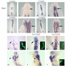
Figure 1
Figure 1
Activation of the Wnt pathway results in increased Pax3 expression in the early ophthalmic trigeminal placode. In situ hybridization showing Pax3 (blue) (A-J) and Fgf8 (red) (F, G). (A-D) Expression of Pax3; (A'-D') are higher magnification images of embryos in (A-D). At 3 somites, Pax3 expression is detectable in the somitic mesoderm, but no transcripts are detected in the neural folds or adjacent ectoderm (A, A', black arrow). At 6 somites, Pax3 transcripts are detectable in the neural folds (B, B'), and transcripts begin to emerge in the cranial ectoderm (black arrow). Pax3 expression becomes more abundant by 8 somites (C, C', black arrow). At 14 somites (HH11+) the Pax3 expressing placode is evident (D, D', black arrow. (E-J) CAGGS Wnt and green fluorescent protein (GFP) are co-electroporated at the midbrain level of the neural folds and targeted to the right side. GFP expression is shown in insets (E', F', G', J'). Embryos were cultured for 6 hours (E, F, G) or 12 hours (H-J). (E) Overexpression of Wnt1 at 6 somites leads to a premature appearance of the earliest Pax3 positive presumptive ophthalmic placodal cells (grey arrow). Embryos injected at the 8 (F) or 10 somite stage (G) also yield a greater number of Pax3 positive cells in the adjacent ectoderm (F, G, compare grey arrow, right, with black arrow, left). No change in isthmic Fgf8 expression (red) is observed (F, G). (H) HH10 embryo electroporated and incubated for 12 hours. The white arrowhead (H) delineates the expanded Pax3 expressing domain compared to the uninjected side (black arrow head). (I, J) Left and right lateral views, respectively, of the embryo viewed dorsally in (H). The arrowheads point to the ophthalmic branch of the trigeminal placode on the injected side (J, white arrowhead), and uninjected side (I, black arrowhead).
Figure 2
Figure 2
Isthmic derived Wnt signals are required at HH8 for maintenance of the early ophthalmic placode identity. In situ hybridization for Pax3 (blue) (B-E, G-J). GFP expression is shown in green (A, F and G). A dominant negative form of Wnt1 (DN Wnt1) was targeted to the right-hand side of the neural tube in all cases. Embryos were co-electroporated with GFP at HH7 (A) or HH10 (F, G) and analyzed 16 and 12 hours later until they reached approximately HH13. When Wnt ligands are perturbed at HH8, expression of Pax3 is reduced in the placodal ectoderm (C, black arrow head; E). The white arrowhead (B) indicates the normally developing placode on the un-injected side. When DN Wnt1 is injected at HH10 Pax3 expression in the placodes is not altered when analyzed at HH13 (G, H, J). The lines in (H) mark the extent through which Pax3 is expressed in the ophthalmic placodes on both sides. Embryos injected with dominant negative Wnt1 at HH8 (I) or HH10 (J) were sectioned to reveal the localization of Pax3 expression. Less Pax3 positive cells were observed in general, and within the mesenchyme, when dominant negative Wnt1 was injected at HH8 (I, right hand side). No obvious differences were observed when dominant negative Wnt1 was electroporated after HH10 (J). Asterisks delineate midbrain (G, H).
Figure 3
Figure 3
Wnt signals act in a temporal manner to regulate Pax3 expression. Explants isolated at HH10 and HH13 were cultured alone (A, C, E) or with SFRP2 (B, D, F) overnight and stained for Pax3 (A-D) and Wnt1 (E-F). Explants cultured with SFRP2 lost the expression of Pax3 (B), compared to controls (A). Cranial ectoderm (ce) isolated at HH13 (C, D) and cultured in the presence of SFRP2 (D) did not lose Islet1 expression compared to controls (C). Midbrain to rhombomere 2 (mb-r2) explants express Wnt1 when cultured alone (E) but lose endogenous Wnt1 transcripts when cultured in the presence of SFRP2 (F) (n = 4/4)
Figure 4
Figure 4
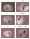
Wnt signals are dependent on FGF signaling for the onset of Pax3 expression and subsequent neuronal differentiation in the cranial ectoderm. Cranial ectoderm explants isolated at the 3 somite stage (as depicted in the cartoon diagram) were grown in collagen gel cultures in the presence of Wnt3A (B, F), FGF8 (C), or Wnt3A and SU5402 (D, G). Explants were stained for the expression of Pax3 (A-D) and Brn3a (E-G). The presence of Wnt3A (B) but not FGF8b (C) was sufficient to induce the onset of Pax3 expression. However, Wnt3A is dependent on FGF signals to initiate Pax3 expression (D). When cranial ectoderm was isolated and cultured for 24 hours until approximately HH16, Wnt3A (F) resulted in expression of Brn3a. Similarly, the activity of Wnt3A was dependent on FGF signals to turn on Brn3a expression (G). Control explants cultured overnight were negative for the expression of Brn3a (E).
Figure 5
Figure 5
Overexpression of Wnt1 leads to ectopic activation of Fgf8 within rhombomere 1 and a hyperdifferentiated trigeminal. Fgf8 expression (blue) detected by in situ hybridization (A-H) and anti-neurofilament (brown) (C-J). Overexpression of Wnt1 at the right-hand side of the neural tube at HH10 results in an expansion of isthmic Fgf8 expression, as analyzed at HH16 (A, B, D, F). (A) Dorsal view (anterior is to the top) of the anterior hindbrain showing expansion of Fgf8 expression in the right neural tube (black arrow). (B) Embryo as in (A) viewed laterally from the right-hand side. Embryos (C-J) are flat mount preparations, anterior is left (C, E, G, I) and right (D, F, H, J). (C) Bisected head at HH16 showing the normal development of the ophthalmic (C, G, white arrows) and maxillomandibular (C, white arrow head) lobes. (D, H) Elevated Wnt signals and expansion of isthmic Fgf8 expression promotes differentiation of both the ophthalmic (black arrows) and maxillomandibular (black arrowhead) branches. When the isthmic organizer (IsO) region is removed (J) and replaced with foil, the trigeminal ganglion appears smaller than the unoperated side (I). (E, F) High magnification images of isthmic domain (C, D). (G, H) High magnification images of ophthalmic lobe in (C, D). The asterisk in (D) delineates the rhombomere 1/2 boundary. The eye (e) is marked for reference in (C, D).
Figure 6
Figure 6
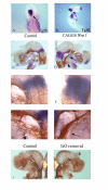
The ophthalmic and maxillomandibular branches of the trigeminal nerve differentiate in response to activated fibroblast growth factor (FGF) signals within the neural tube. In situ hybridization detects Islet1 expression (A, C-D, E-J), green fluorescent protein (GFP) expression (B, G', J') and anti-Islet1 (K, L) expression. FGF8, Wnt1 or FGF8 and Wnt1 were electroporated at the right side of the neural tube at HH10, and embryos were cultured until HH16 (A-L). Overexpression of FGF8 results in premature differentiation of the ophthalmic and maxillomandibular branches (compare white arrow head in (D) with the black arrow head in (C)). Similarly, following electroporation of Wnt1 in the neural tube, the expression of Islet1 is more pronounced in the right trigeminal placodes (E, G, white arrowheads) compared to the untargeted side (F, white arrowhead). Overexpression of both Wnt1 and FGF8 together (H, I, J, black arrowheads) also results in similar expansion of Islet1 expression. Confocal analysis for Islet1 expression (red) within the developing placodes reveals that both the ophthalmic and maxillomandibular branches appear prematurely differentiated (L) compared to the un-targeted side (K). (C, D) High magnification images of flat mount in (A); (F, G) high magnification images of flat mount in (E); (I, J) higher magnification images of flat mount in (H).
Figure 7
Figure 7
Addition of fibroblast growth factor (FGF) or Wnt3A results in a similar expression of Islet1 in the ectoderm compared to the addition of Wnt3A and FGF8 together. In situ hybridization of Islet1 (blue) (A-F) of explants grown over night in culture. Explants in (A, C, E) were left untreated. Treated explants were grown in the presence of FGF8 (B), Wnt3A (D) and Wnt3A and FGF8 (F). FGF8 (B) or Wnt3A (D) addition resulted in a larger domain of Islet1 expression compared to controls (A, C). Addition of both Wnt3A and FGF8 (F) did not result in an increase in the domain of Islet1 expression compared to either ligand alone.
Figure 8
Figure 8
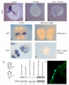
Fibroblast growth factor (FGF) signaling via the mitogen-activated protein kinase (MAPK) pathway is required for the maintenance of Islet1 expression in the trigeminal placodes. Cranial ectoderm explants (A-C, F, G) and midbrain to rhombomere 2 (mb-r2) (D, E) were excised at HH10 and grown overnight until approximately HH16. In situ hybridizations for Islet1 (A-C, F, G) and Fgf8 (D-E) expression are shown in blue. Explants were cultured in SU5402 (B), Wnt3A and SU5402 (C) or PD184352 (E, G). The presence of SU5402 resulted in the loss of Islet1 expression (B) compared to controls (A). In the presence of SU5402, Wnt3A did not maintain Islet1 expression (C). The presence of a selective MEK antagonist (PD184352, 2 nM) led to a loss of Fgf8 expression in mb-r2 explants (E) compared to controls (D). Inhibition of MEK activity also resulted in a complete loss of Islet1 expression (G) compared to controls (F). (D-G), control explants (left ce) and experimental explants (right ce) were isolated from the same embryo. (H) Western blots of HH10 mb-r2 explants (lanes 1, 3 and 5) and HH10 cranial ectoderm (ce; lanes 2, 4 and 6). Mb-r2 and cranial ectoderm explants lose double phosphorylated ERK1/2 (dpERK1/2) activity in the presence of 50 μM SU5402 (lanes 3 and 4). In all other lanes, isolated tissue is positive for dpErk1/2 at HH10 (lanes 1 and 2), and also for nuclear β-catenin (lanes 5 and 6). Actin is shown as a loading control. (I) Immunostain showing the expression of dpERK in the cranial ectoderm.

References

    1. D'Amico-Martel A, Noden DM. Contributions of placodal and neural crest cells to avian cranial peripheral ganglia. Am J Anat. 1983;166:445–468. doi: 10.1002/aja.1001660406. - DOI - PubMed
    1. Ayer-Le Lievre CS, Le Douarin NM. The early development of cranial sensory ganglia and the potentialities of their component cells studied in quail-chick chimeras. Dev Biol. 1982;94:291–310. doi: 10.1016/0012-1606(82)90349-9. - DOI - PubMed
    1. Northcutt RG, Brandle K. Development of branchiomeric and lateral line nerves in the axolotl. J Comp Neurol. 1995;355:427–454. doi: 10.1002/cne.903550309. - DOI - PubMed
    1. Schlosser G. Induction and specification of cranial placodes. Dev Biol. 2006;294:303–351. doi: 10.1016/j.ydbio.2006.03.009. - DOI - PubMed
    1. Stark MR, Sechrist J, Bronner-Fraser M, Marcelle C. Neural tube-ectoderm interactions are required for trigeminal placode formation. Development. 1997;124:4287–4295. - PubMed

Publication types

MeSH terms

Substances

LinkOut - more resources