. 2008 Nov-Dec;2(6):269-79.
doi: 10.4161/fly.7352. Epub 2008 Nov 5.

The role of the RING-finger protein Elfless in Drosophila spermatogenesis and apoptosis

Affiliations

The role of the RING-finger protein Elfless in Drosophila spermatogenesis and apoptosis

Jason C Caldwell et al. Fly (Austin). 2008 Nov-Dec.

Abstract

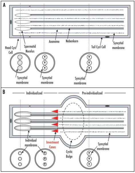
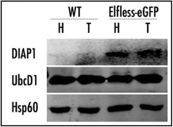
elfless (CG15150, FBgn0032660) maps to polytene region 36DE 5' (left) of reduced ocelli/Pray for Elves (PFE) on chromosome 2L and is predicted to encode a 187 amino acid RING finger E3 ubiquitin ligase that is putatively involved in programmed cell death (PCD, e.g., apoptosis). Several experimental approaches were used to characterize CG15150/elfless and test whether defects in this gene underlie the male sterile phenotype associated with overlapping chromosomal deficiencies of region 36DE. elfless expression is greatly enhanced in the testes and the expression pattern of UAS-elfless-EGFP driven by elfless-Gal4 is restricted to the tail cyst cell nuclei of the testes. Despite this, elfless transgenes failed to rescue the male sterile phenotype in Df/Df flies. Furthermore, null alleles of elfless, generated either by imprecise excision of an upstream P-element or by FLP-FRT deletion between two flanking piggyBac elements, are fertile. In a gain-of-function setting in the eye, we found that elfless genetically interacts with key members of the apoptotic pathway including the initiator caspase Dronc and the ubiquitin conjugating enzyme UbcD1. DIAP1, but not UbcD1, protein levels are increased in heads of flies expressing Elfless-EGFP in the eye, and in testes of flies expressing elfless-Gal4 driven Elfless-EGFP. Based on these findings, we speculate that Elfless may regulate tail cyst cell degradation to provide an advantageous, though not essential, function in the testis.


Figures

Figure 1
Figure 1
Spermatid individualization. (A) Following elongation of the axoneme, mitochondria and the tail cyst cell, the head cyst cell-associated spermatid nuclei condense into needle-like morphologies. For clarity, only three of the 64 spermatid units are shown. The spermatids are still connected by a syncytial membrane (jagged lines). The vertical dashed lines indicate areas diagrammed in cross-section below. The interconnected syncyctial membrane surrounding the various spermatid structures is shown (thick black incomplete circles). (B) The investment cones (triangles) travel the length of the spermatids to (1) individualize each member of the 64-unit bundle in its own plasma membrane and (2) remove the shared, syncytial cytoplasm. These cones have already individualized the spermatid nuclei and some of the axoneme/nebenkern. As the investment cones travel caudally (rightward), the cytoplasmic waste is collected and forms a so-called cystic bulge. At the end of individualization the waste bag is removed (not shown). In (B), the individualized areas are indicated by thick black circles representing membranes tightly surrounding the spermatid structures while the pre-individualized areas are indicated by thick black incomplete circles.
Figure 2
Figure 2
The btv-male sterile-rdo overlapping deficiency region. (A) Representative squash of Df/Df testes showing amotile sperm that remain encysted. (B) The genetic map of the btv-male sterile-rdo region derived from overlapping deficiencies TW119 and TW201. In the flanking regions uncovered by only one of the deficiencies, only some loci are listed. (C) A subregion of the larger Df/Df including elfless and three other small predicted genes (btv and CG5674 are not shown at this magnification). Arrows indicate the direction of transcription. Positions of transposable element insertions used in this study are shown by grey triangles. (D) The elfless intronexon structure shows the three exons (boxes) connected by two introns (thin lines). The 5′UTR (black box) and 3′UTR (grey box) are also indicated. (E) The predicted domain structure of the elfless protein contains a nuclear localization signal (NLS; green box) and C-terminal RING domain (blue box).
Figure 3
Figure 3
The elfless nucleotide and amino acid sequences. The elfless 5′UTR is shown in blue, exons are in black (ATG start codon in bold), introns in grey and the 3′UTR in red with putative polyadenylation site (AATAAA) in bold. The elfless protein consists of a C3HC4-type RING domain (blue shaded box region with consensus Cys and His residues indicated in bold) and a nuclear localization signal (green box). These sequences are based on the AT24563 cDNA clone structure.
Figure 4
Figure 4
elfless transcripts are greatly enhanced in testes. Levels of elfless transcripts, isolated from various tissues, relative to Gapdh2 control transcripts. elfless is most abundant in testes (∼1 copy of elfless for every 3 copies of Gapdh2), and is less abundant in whole males. elfless is nearly undetectable in carcasses (males with testes removed) and females. elfless expression in testes is significantly greater than all other tissues (p < 0.001 ANOVA/Tukey) and likewise elfless transcript levels in whole males is greater than carcasses and females (***p < 0.001 ANOVA/Tukey). elfless expression levels do not distinguishably differ between carcasses and females.
Figure 5
Figure 5
elfless is expressed in testes. All UAS-constructs are driven by elfless-Gal4 and the expression pattern is restricted to the testes. ((A–C), UAS-elfless-EGFP; (D–F) UAS-cytoplasmic-GFP) (A) UAS-elfless-EGFP expression is limited to the tail cyst cell nuclei, confirming the predicted nuclear localization signal for elfless. (B) nucDsRed co-localizes with elfless-EGFP in the tail cyst cell nuclei and also exhibits significant spurious expression (independent of Gal4 driver) in the spermatid and head cyst cell nuclei (non co-localized regions in C). (C) Merge of (A and B). (D) UAS-cytoplasmic-GFP expression (without nuclear localization signal) is seen throughout the tail cyst cell. (E) DIC (F) Merge of (D and E). Scale bars = 150 μm.
Figure 6
Figure 6
Elfless-EGFP is co-expressed with a tail cyst cell marker. A testis from a male with elfless-Gal4 driving UAS-elfless-EGFP in the presence of the tail cyst cell enhancer trap P{lacW}498 was processed for X-gal staining to reveal the tail cyst cell nuclei (A and A′). Sufficient EGFP fluorescence remained in these testes for imaging despite some increased background (B and B′). Imaging both simultaneously (C and C′) shows significant co-incidence of Elfless-EGFP and enhancer trap expression. The red box in (A) indicates the region shown at higher magnification in (A′–C′).
Figure 7
Figure 7
Ectopic mis-expression of elfless induces apoptosis. (A, C and E) Light microscopy (B, D and F) SEM with insets. (A and B) w; GMR-Gal4/+; UAS-elfless/+ flies exhibit pigment loss and other defects such as loss or duplication of the interommatidial bristles and often the eye surface morphology is irregular. (C and D) w; GMR-Gal4/+; UAS-elfless/ubcD1 flies exhibit almost complete pigment loss and severe bristle and eye shape/size phenotypes and this genetic combination shows reduced viability. (E and F) w; GMR-Gal4/+; UAS-elfless/Dronc flies exhibit less severe pigment and morphological defects than w; GMR-Gal4/+; UAS-elfless/+ alone.
Figure 8
Figure 8
DIAP1, but not UbcD1, is increased upon Elfless expression. Western blot of protein isolated from GMR-Gal4 control heads (H), or Elfless-Gal4 control testes (T), was probed with antibodies against DIAP1, UbcD1 or the loading control, Hsp60. Compared to these controls (labeled WT), protein extracts of the corresponding tissues from their siblings, which also carry the UAS-elfless-EGFP construct (labeled Elfless-eGFP), show increased levels of DIAP1, but no change in UbcD1 levels is evident.
Figure 9
Figure 9
Model for elfless in apoptosis. In these simplified models of apoptosis, the strength of inhibition (t-bars), activation (arrowheads) or ubiquitination (inverted arrowheads) is represented by line weight. (A) In a wild type non-apoptotic cell, the key regulatory molecule is DIAP1. This protein inhibits the downstream activator caspase, Dronc, and the effector caspases (Drice and Dcp-1). Likewise, Dronc is a target of ubcD1-dependent ubiquitination. (B) When a wild type cell receives an intrinsic or extrinsic signal to die, the pro-apoptotic genes, Grim-Rpr-Hid inhibit DIAP1 and DIAP1 auto-ubiquitinates in a ubcD1-dependent manner. The downstream caspases are no longer strongly inhibited and the cell becomes apoptotic. In this genetic background, two copies of ubcD1 are present and therefore a significant level of ubiquitination of Dronc is evident and this balance prevents “excessive” cell death. (C) In this model for the role elfless as a direct or indirect (black box) negative regulator of ubcD1 activity. When elfless is mis-expressed with GMR-Gal4 ubcD1 is strongly inhibited. This presumably results in strong suppression of caspsases by DIAP1, but since ubcD1 also acts further downstream, the increased apoptosis is attributable to mis-regulation of Dronc by downregulation of ubcD1 by elfless. Consistent with this model, this effect is exacerbated in ubcD1 heterozygotes and is attenuated in Dronc heterozygotes.

Similar articles

Cited by

References

    1. Castrillon DH, Gönczy P, Alexander S, Rawson R, Eberhart CG, Viswanathan S, DiNardo S, Wasserman SA. Toward a molecular genetic analysis of spermatogenesis in Drosophila melanogaster: characterization of male-sterile mutants generated by single P element mutagenesis. Genetics. 1993;135:489–505. - PMC - PubMed
    1. Gönczy P, Viswanathan S, DiNardo S. Probing spermatogenesis in Drosophila with P-element enhancer detectors. Development. 1992;114:89–98. - PubMed
    1. Gönczy P, DiNardo S. The germ line regulates somatic cyst cell proliferation and fate during Drosophila spermatogenesis. Development. 1996;122:2437–47. - PubMed
    1. Fuller MT. Spermatogenesis. In: Bate M, Martinez Arias A, editors. The Development of Drosophila. Cold Spring Harbor Press; Cold Spring Harbor: 1993. pp. 71–147.
    1. Williamson A, Lehmann R. Germ cell development in Drosophila. Annu Rev Cell Dev Biol. 1996;12:365–91. - PubMed

Publication types

MeSH terms

LinkOut - more resources