. 2008 Nov-Dec;2(6):306-9.
doi: 10.4161/fly.7427. Epub 2008 Nov 15.

Drosophila embryonic 'fibroblasts': extending mutant analysis in vitro

Affiliations

Drosophila embryonic 'fibroblasts': extending mutant analysis in vitro

Amanda A Simcox et al. Fly (Austin). 2008 Nov-Dec.

Abstract

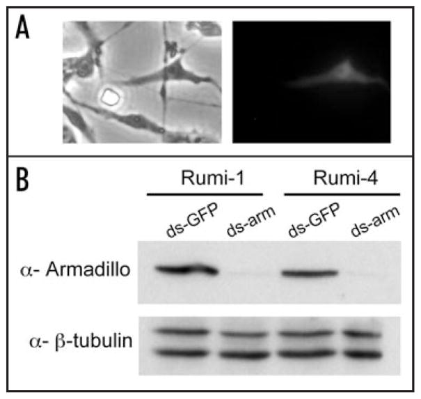
The in vivo analysis of Drosophila using genetics, with almost a hundred year history, has produced an immense body of knowledge about biology. In vitro analysis, while arguably the poor cousin to its in vivo relative, has a utility--in biochemical analyses and in cell-based screening, for example, with RNAi. A major block to the development of in vitro analysis has been the lack of an efficient genetic method to derive cell lines from mutant Drosophila strains. We recently discovered that expression of activated Ras (Ras(V12)) provides cells in vitro with both a survival and a proliferative advantage and hence promotes the generation of cell lines. In this addendum, we provide new data describing the genesis of seven cell lines corresponding to a rumi mutant, which demonstrate that the method can be used to derive lines and study genetic mutants in vitro.


Figures

Figure 1
Figure 1
Genetic cross to produce rumi26 mutant embryos expressing activated Ras (RasV12). Primary cultures were established from a mixture of embryos of all four genotypes. The boxed embryos are the desired genotype. The UAS-RasV12 insertion site was mapped to 3R:7,394,981 (it is accompanied by a 151 bp deletion) within the 5′ region of CG14709. The Act-GAL4 insertion site was mapped to 3R:25,584,989 between CG1973 and kayak. The Act5C-GAL4 transgene is inserted in sequence corresponding to a transposable element and is currently being mapped further genetically.
Figure 2
Figure 2
Landmarks in passaging history. The plot shows when each line (Rumi 1–7) was passaged starting at the time the primary culture was confluent (Passage 1). The primary cultures were confluent in about three weeks (line 4 took longer because the primary culture was sparse). Cells in confluent cultures were harvested and half were seeded into a new flask (1 in 2 dilution). The number of days between passages was more variable in early passages (P1–10) than in later passages. All lines have now been passaged 24 or more times and the time between passages is 5–12 days. In the lines that have now been passaged 25 or more times the cells can be split at higher dilutions (1 in 3 or 1 in 4).
Figure 3
Figure 3
Evolution of primary cultures to cell lines of spindle-shaped cells. (A) Examples of cell types from primary cultures. (B) In early passages, spindle-shaped cells are the prevalent type. (C) Established Rumi cell lines and a control (Ras7) cell line are comprised of similar spindle-shaped cells. (P, passage; all panels are phase contrast images at the same magnification).
Figure 4
Figure 4
Cell lines are deficient for Rumi. Diagnostic PCR tests and Western blotting were used to genotype cells in the Rumi lines and determine their protein expression levels. Together the results show that 6/7 Rumi lines were pure populations of rumi26 homozygous cells. One line had a fraction of non-mutant cells but this fraction is assumed to be low because the cells had undetectable levels of Rumi protein. (A) Control (Ras7) cells have a diagnostic band corresponding to the wild-type rumi gene (1832 bp), whereas, each of the 7 Rumi lines (R-1 through R-7) have a 344 bp band diagnostic for the rumi26 deletion. This shows that most cells in the Rumi lines are homozygous for rumi26. (B) In order to detect rare non-homozygous mutant cells, another PCR test was performed using primers that only amplify the wild-type gene. Both control Ras7 and Rumi line 3 cells had the expected 615 bp fragment. This shows that some fraction of cells in Rumi line-3 have a wild type rumi gene. (C) Western blotting was used to analyze Rumi protein in control (Ras7) and Rumi line 3 and 6 cells (R3 and R6). Only the control cells expressed detectable levels of Rumi protein. Actin was used as a control to show that similar levels of protein were loaded.
Figure 5
Figure 5
Rumi cells can be transfected and are susceptible to RNAi. (A) The left panel shows a phase contrast image and the right panel shows a fluorescent image. One Rumi line-5 cell in the field is expressing MAPK-YFP. Cells were transfected with an Act5C-ERK-YFP plasmid using Effectene (Qiagen) and assayed for GFP expression 48 hours after transfection. (B) Western blot of Rumi-1 and Rumi-4 cells treated with either a control ds-GFP RNA or ds-arm RNA. The ds-arm RNA substantially reduces the level of Armadillo protein in cells from both cell lines. β-tubulin levels show similar amounts of proteins were loaded. PCR products corresponding to armadillio and GFP were used in an in vitro transcription reaction (Megascript, Ambion) to generate double stranded RNA. Rumi cells were seeded in 6-well plates 24 hrs prior to transfection with 30 μg dsRNA using Effectene (Qiagen). Cells were assayed after 48 hrs by Western blotting with monoclonal antibodies against Armadillo (1/100, N2 7A1, Developmental Studies Hybridoma Bank) and polyclonal antibodies against β-tubulin (1/2000 SC33749, Santa Cruz Biotechnology).

References

    1. Simcox A, Mitra S, Truesdell S, Paul L, Chen T, Butchar JP, Justiniano S. Efficient genetic method for establishing Drosophila cell lines unlocks the potential to create lines of specific genotypes. PLoS Genet. 2008;4:1000142. - PMC - PubMed
    1. Schneider I. Cell lines derived from late embryonic stages of Drosophila melanogaster. J Embryol Exp Morphol. 1972;27:353–65. - PubMed
    1. Echalier G, Ohanessian A. In vitro culture of Drosophila melanogaster embryonic cells. In Vitro. 1970;6:162–72. - PubMed
    1. Echalier G. Drosophila cells in culture. New York: Academic Press; 1997.
    1. Acar M, Jafar-Nejad H, Takeuchi H, Rajan A, Ibrani D, Rana NA, Pan H, Haltiwanger RS, Bellen HJ. Rumi is a CAP10 domain glycosyltransferase that modifies Notch and is required for Notch signaling. Cell. 2008;132:247–58. - PMC - PubMed

Publication types

LinkOut - more resources