. 2009 Jan;19(1):87-9.
doi: 10.1097/FPC.0b013e32830fbde4.

Identification of a novel haplotype of the human catechol-O-methyltransferase gene

Affiliations

Identification of a novel haplotype of the human catechol-O-methyltransferase gene

Hyoung-Woo Bai et al. Pharmacogenet Genomics. 2009 Jan.

Abstract

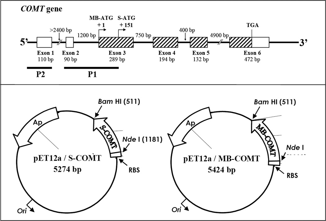
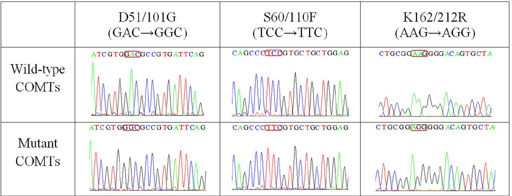
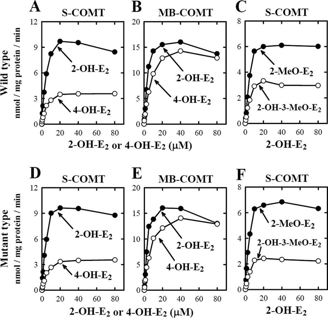
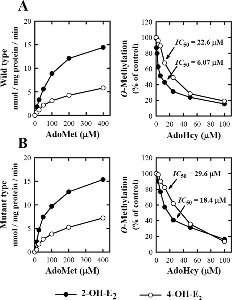
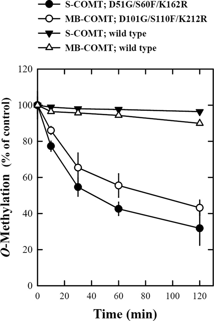
Human catechol-O-methyltransferase (COMT; EC 2.1.1.6) catalyzes the transfer of the methyl group to a variety of endogenous and exogenous catechol substrates using S-adenosyl-L-methionine as the methyl donor. This enzymatic O-methylation plays an important role in the inactivation of biologically active and toxic catechols. A number of studies in recent years have sought to characterize the polymorphism of human COMTs and also to determine the catalytic activity of polymorphic enzymes. We report here the identification of a new haplotype of the human COMT gene with triplet point mutations, which encodes the D51G/S60F/K162R mutant of the soluble COMT and the D101G/S110F/K212R mutant of the membrane-bound COMT. Kinetic analysis showed that these new COMT variants had essentially the same kinetic characteristics and catalytic activity as the wild-type COMTs for the O-methylation of 2-hydroxyestradiol and 4-hydroxyestradiol in vitro, but they have a significantly reduced thermostability at 37 degrees C.


Figures

Figure 1
Figure 1
Upper panel: Structure of the human COMT gene. The boxes represent exons and the thin lines between the boxes represent introns. The hatched boxes indicate protein-coding regions. The size of each exon and intron is as indicated. The positions of the initiation codons for transcription of S-COMT and MB-COMT mRNAs are indicated as S-ATG and MB-ATG. The two known promoters, P1 and P2, are shown by black bars. Note that the P1 promoter for transcritpion of S-COMT overlaps with the initiation codon and part of the coding sequence for MB-COMT. Lower panel: Construction of the pET12a/S-COMT and pET12a/MB-COMT expression vectors based on the vector pET12a. The S-COMT or MB-COMT cDNA was cloned into the NdeI and BamH I sites of pET12a to form the pET12a/S-COMT or pET12a/MB-COMT expression vectors. Each of the expression vectors was under the control of the T7 promoter and lacO-operator. Expression was induced by addition of 0.5 mM isopropylthio-β-D-galactoside.
Figure 2
Figure 2
Sequencing results of the wild-type and mutant human S- and MB-COMTs.
Figure 3
Figure 3
Relationship between catechol estrogen concentrations and their rate of O-methylation by wild-type human COMTs (A, B and C) and mutant human COMTs (D, E, and F). The upper right panel (C) showed the rate of 2-O-methylation and 3-O-methylation of 2-OH-E2 catalyzed by the wild-type S-COMT, and the lower right panel (F) showed the rate of 2-O-methylation and 3-O-methylation of 2-OH-E2 by mutant S-COMT. The incubation mixture consisted of 10 different concentrations (0, 0.31, 0.63, 1.25, 2.5, 5, 10, 20, 40, and 80 µM) of each substrate, 1.2 mM MgCl2, 100 µM AdoMet (containing 0.5 mCi [methyl-3H]AdoMet), 1 mM 1,4-dithiothreitol, and the recombinant COMT protein (at 16.2 µg/mL for S-COMT or 17.1 µg/mL for MB-COMT). The incubations were carried out at 37°C for 15 minutes. Note that the rate of its total methylation was based on liquid scintillation counting of the radioactivity extracted with ethyl acetate. The rates for its 2-O- and 3-O-methylation (C and F) were determined by using HPLC that separately quantified the amount of 2-methoxyestradiol (2-MeO-E2) and 2-OH-E2 3-methyl ether (2-OH-3-MeO-E2) formed. Each value is the mean of duplicate measurements.
Figure 4
Figure 4
Biochemical properties of the in vitro O-methylation of catechol substrates with respect to AdoMet and AdoHcy concentrations. The representative substrates used in these assays were 2-OH-E2 and 4-OH-E2 at 10 µM concentration. The incubation mixture consisted of the substrate, 100 µM [methyl-3H]AdoMet (containing 0.2 µCi or as indicated), 16.2 µg/mL of wild-type S-COMT (A) or mutant S-COMT (B), 1 mM 1,4-dithiothreitol, and 1.2 mM MgCl2 in a final volume of 300 µL Tris-HCl buffer (50 mM) at pH 7.4. The incubations were carried out at 37°C for 15 minutes. Each value is the mean of duplicate measurements.
Figure 5
Figure 5
Stabilities of the wild-type and mutant human recombinant S- and MB-COMTs. The reaction mixtures consisted of the recombinant COMT protein (at 16.2 µg/mL for S-COMT or 17.1 µg/mL for MB-COMT), 1.2 mM MgCl2, 100 µM AdoMet (containing 0.5–1 mCi [methyl-3H]AdoMet), 2-OH-E2 as a substrate and 1 mM 1,4-dithiothreitol in Tris-HCl buffer (50 mM, pH 7.4). The final volume of the reaction mixture was usually 300 µL. The reaction was initiated by addition of the recombinant human COMT protein and carried out at 37°C for 15 minutes. To test the thermostability of the mutant and wild-type COMTs, the enzymes were first preincubated at 37°C for the indicated length of time immediately before testing their catalytic activity for the O-methylation of 2-OH-E2 (at 10 µM). Each value is the mean ± S.D. of triplicate measurements.
Figure 6
Figure 6
The wild-type human S-COMT (A), the D51G/S60F/K162R mutant S-COMT built according to the homology model of the wild-type human S-COMT (B) or according to the crystallographic structure of the rat S-COMT (PDB code: 1VID) (C). The figure is drawn with the PyMOL software. Secondary structures are shown with colored ribbons with blue for N-terminus and red for C-terminus. AdoMet, Mg2+ and substrates are not included in this model. The amino acids at mutation sites (D51, S60 and K162 for wild-type S-COMT and G51, F60 and R162 for mutant S-COMT) are shown in white sticks. Hydrogens are omitted from the amino acids.
Figure 7
Figure 7
The wild-type human S-COMT (A) and the D51G/S60F/K162R mutant S-COMT built according to the homology model of the wild-type human S-COMT (B). The enzymes are complexed with 2-OH-E2 in the catalytic pocket in its geometry for 2-O-methylation. The figure is drawn with the PyMOL software. Secondary structures are shown with colored ribbons with blue for N-terminus and red for C-terminus. AdoMet is colored red, 2-OH-E2 is colored green and Mg2+ is colored magenta. The amino acids at mutation sites (D51, S60 and K162 for wild-type S-COMT and G51, F60 and R162 for mutant S-COMT) are shown in white sticks. Hydrogens are omitted from AdoMet, 2-OH-E2 and the amino acids.

Similar articles

References

    1. Axelrod J, Tomchick R. Enzymatic O-methylation of epinephrine and other catechols. J Biol Chem. 1958;233:702–705. - PubMed
    1. Axelrod J. Methylation reactions in the formation and metabolism of catecholamines and other biogenic amines. Pharmacol Rev. 1996;18:95–113. - PubMed
    1. Zhu BT. Catechol-O-Methyltransferase (COMT)-mediated methylation metabolism of endogenous bioactive catechols and modulation by endobiotics and xenobiotics: importance in pathophysiology and pathogenesis. Curr Drug Metab. 2002;3:321–349. - PubMed
    1. Tenhunen J, Salminen M, Jalanko A, Ukkonen S, Ulmanen I. Structure of the rat catechol-O-methyltransferase gene: separate promoters are used to produce mRNAs for soluble and membrane-bound forms of the enzyme. DNA Cell Biol. 1993;12:253–263. - PubMed
    1. Zhu BT, Liehr JG. Quercetin increases the severity of estradiol-induced tumorigenesis in hamster kidney. Toxicol Appl Pharmacol. 1994;125:149–158. - PubMed

Publication types