Review
. 2009 Jan;15(1):4-11.
doi: 10.1097/MCP.0b013e32831da8d3.

Mucus hypersecretion in asthma: causes and effects

Affiliations
Review

Mucus hypersecretion in asthma: causes and effects

Christopher M Evans et al. Curr Opin Pulm Med. 2009 Jan.

Abstract

Purpose of review: Airway mucus plugging has long been recognized as a principal cause of death in asthma. However, molecular mechanisms of mucin overproduction and secretion have not been understood until recently. These mechanisms are reviewed together with ongoing investigations relating them to lung pathophysiology.

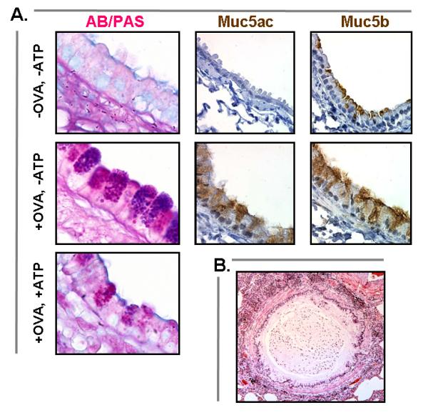
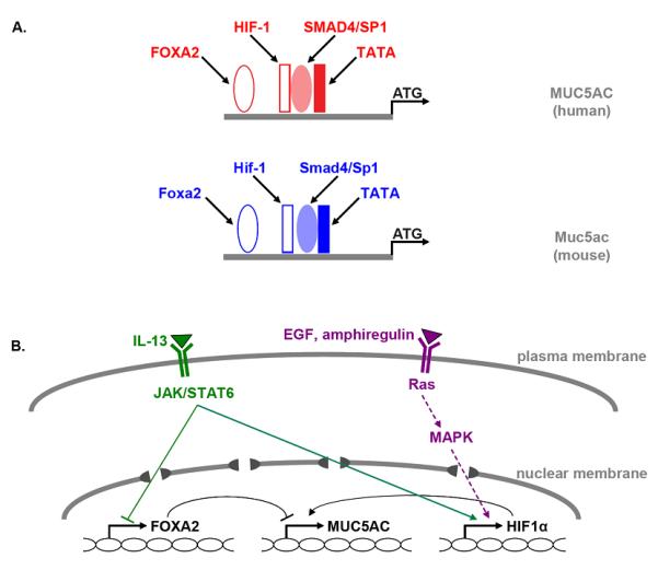
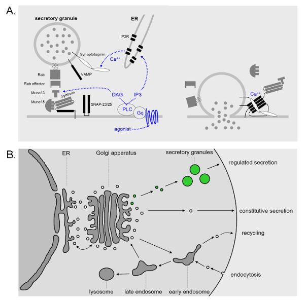
Recent findings: Of the five secreted gel-forming mucins in mammals, only MUC5AC and MUC5B are produced in significant quantities in intrapulmonary airways. MUC5B is the principal gel-forming mucin at baseline in small airways of humans and mice, and therefore likely performs most homeostatic clearance functions. MUC5AC is the principal gel-forming mucin upregulated in airway inflammation and is under negative control by forkhead box a2 (Foxa2) and positive control by hypoxia inducible factor-1 (HIF-1). Mucin secretion is regulated separately from production, principally by extracellular triphosphate nucleotides that bind P2Y2 receptors on the lumenal surface of airway secretory cells, generating intracellular second messengers that activate the exocytic proteins, Munc13-2 and synaptotagmin-2.

Summary: Markedly upregulated production of MUC5AC together with stimulated secretion leads to airflow obstruction in asthma. As MUC5B appears to mediate homeostatic functions, it may be possible to selectively inhibit MUC5AC production without impairing airway function. The precise roles of mucin hypersecretion in asthma symptoms such as dyspnea and cough and in physiologic phenomena such as airway hyperresponsiveness remain to be defined.


Figures

Figure 1
Figure 1
Mucus hypersecretion gr1 (a) In ovalbumin-sensitized and challenged mice (+OVA, -ATP), there is a dramatic increase in the number of AB—PAS-positive mucous cells in the tracheobronchial airways (d). This increase is apparent 24h after challenge and peaks at 3–7 days. Despite the lack of AB—PAS staining at baseline (a), intracellular Muc5b (c) can be demonstrated by sensitive immunohistochemical techniques at the cell apex (-OVA, -ATP). It becomes redistributed throughout the distended cytoplasm (f) during mucous metaplasia (+OVA, -ATP). Muc5ac is not apparent immunohistochemically at baseline (b) but is strongly expressed (f) during mucous metaplasia (+OVA, -ATP). After stimulation of metaplastic mucous cells with aerosolized ATP, there is rapid secretion of most of the accumulated intracellular mucin (+OVA, +ATP) [10,11•]. (b) Airway from a patient who died from asthma showing extensive infiltration of the airway wall and surrounding lung tissue with inflammatory cells and mucus filling the airway lumen. AB—PAS, alcian blue and/or periodic acid Shiff; OVA, ovalbumin. Reproduced with permission from [10,11•] and by courtesy of Martha Warnock, University of California at San Francisco.
Figure 2
Figure 2
Transcriptional control of Muc5ac production gr2 (a) Conserved consensus-binding sites for transcription factors in the core promoters of the human MUC5AC (red) and the mouse Muc5ac (blue) genes are indicated. (b) Known pathways for activation of MUC5AC/Muc5ac gene transcription by IL-13 (green) and EGFR ligands (violet) are illustrated. Solid lines indicate direct protein interaction with target gene (i.e., STAT6), dotted lines indicate multiple steps of interaction (i.e., EGFR pathway), arrowheads indicate positive interaction, and bars indicate inhibitory interactions (see text for citations). EGF, epidermal growth factor; EGFR, epidermal growth factor receptor; Foxa2, forkhead box a2; HIF, hypoxia inducible factor; JAK, janus-activated kinase; MAPK, mitogen-activated protein kinase; STAT, signal transducer and activator of transcription.
Figure 3
Figure 3
Mechanism of polymeric mucin secretion gr3 (a) Extracellular nucleotides in the airway surface liquid layer bind to apical P2Y2 receptors that activate the trimeric G-protein Gq, which in turn activates phospholipase C β1, generating the intracellular second messengers, diacylglycerol and inositol trisphosphate (IP3) [13•]. Diacylglycerol directly induces mucin granule exocytosis by activating the priming protein, Munc13-2 [12••], and indirectly regulates exocytosis by activating protein kinase Cε [37•]. IP3 induces the release of Ca2+ from intracellular stores [38,39•,40,41], resulting in a rise in cytoplasmic Ca2+ that rapidly triggers mucin granule exocytosis through the activation of synaptotagmin-2. In airway secretory cells, IP3 receptors are localized to endoplasmic reticulum that lies in close apposition to mucin granules at the apical pole (C. William Davis, personal communication). The reliance of airway secretory cells on intracellular Ca2+ stores to activate exocytosis may reflect the instability of Ca2+ concentrations in airway lining fluid that is directly exposed to the external environment [42], in contrast to nonexocrine secretory cells bathed in interstitial fluid or plasma with tightly controlled Ca2+ concentrations. Activation of Munc13 and synaptotagmin allows formation of a four-helix bundle termed the core complex (black rectangles) that draws secretory granule and plasma membranes tightly together and induces their fusion (right). This leads to the release of granule contents, including polymeric mucins, into the airway lumen. The molecular identities of the core complex isoforms in airway secretory cells are not yet known. (b) Data from mouse genetic models suggest that both baseline and stimulated mucin secretion occur through varying rates of activity of a single regulated exocytic machinery acting on a single population of mucin secretory granules (green). DAG, diacylglycerol; ER, endoplasmic reticulum; IP3R, inositol trisphosphate receptor; PLC, phospholipase C; VAMP, vesicle-associated membrane protein.

References

    1. Thornton DJ, Sheehan JK. From mucins to mucus: toward a more coherent understanding of this essential barrier. Proc Am Thorac Soc. 2004;1:54–61. - PubMed
    1. Thornton DJ, Rousseau K, McGuckin MA. Structure and function of the polymeric mucins in airways mucus. Annu Rev Physiol. 2008;70:459–486. An authoritative, up-to-date review of the gel-forming mucins of the airway.

    1. Williams OW, Sharafkhaneh A, Kim V, et al. Airway mucus: from production to secretion. Am J Respir Cell Mol Biol. 2006;34:527–536. - PMC - PubMed
    1. Evans CM, Koo JS. Airway mucus: the good, the bad, the sticky. Pharmacol Ther. 2008 (in press) - PMC - PubMed
    1. Knowles MR, Boucher RC. Mucus clearance as a primary innate defense mechanism for mammalian airways. J Clin Invest. 2002;109:571–577. - PMC - PubMed

Publication types