. 2009 Jan 22;457(7228):490-4.
doi: 10.1038/nature07547. Epub 2008 Dec 10.

Endochondral ossification is required for haematopoietic stem-cell niche formation

Affiliations

Endochondral ossification is required for haematopoietic stem-cell niche formation

Charles K F Chan et al. Nature. .

Abstract

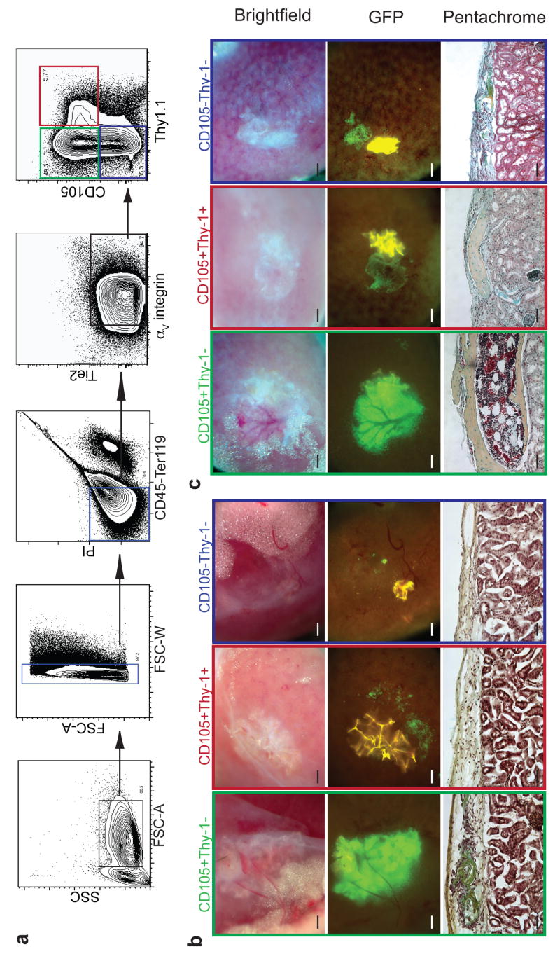
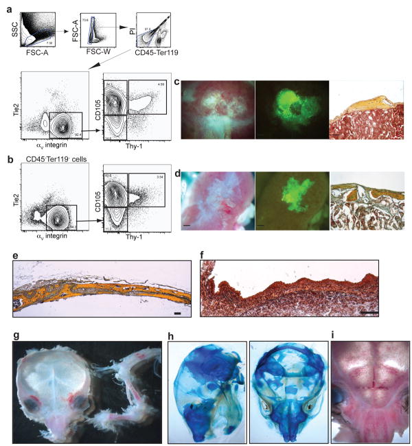
Little is known about the formation of niches, local micro-environments required for stem-cell maintenance. Here we develop an in vivo assay for adult haematopoietic stem-cell (HSC) niche formation. With this assay, we identified a population of progenitor cells with surface markers CD45(-)Tie2(-)alpha(V)(+)CD105(+)Thy1.1(-) (CD105(+)Thy1(-)) that, when sorted from 15.5 days post-coitum fetal bones and transplanted under the adult mouse kidney capsule, could recruit host-derived blood vessels, produce donor-derived ectopic bones through a cartilage intermediate and generate a marrow cavity populated by host-derived long-term reconstituting HSC (LT-HSC). In contrast, CD45(-)Tie2(-)alpha(V)(+)CD105(+)Thy1(+) (CD105(+)Thy1(+)) fetal bone progenitors form bone that does not contain a marrow cavity. Suppressing expression of factors involved in endochondral ossification, such as osterix and vascular endothelial growth factor (VEGF), inhibited niche generation. CD105(+)Thy1(-) progenitor populations derived from regions of the fetal mandible or calvaria that do not undergo endochondral ossification formed only bone without marrow in our assay. Collectively, our data implicate endochondral ossification, bone formation that proceeds through a cartilage intermediate, as a requirement for adult HSC niche formation.


Figures

Figure 1
Figure 1
Fetal bone (fb) cells can initiate an ectopic HSC niche. a, Ectopic bone formed by GFP-labelled, 14.5 dpc fb cells 32 days after subrenal capsule transplant (scale bar = 500 μM). b, Representative section of graft stained with pentachrome (yellow=osteoid, greenish blue= cartilage), cartilaginous region in black circle (scale bar = 100 μM). c, Safranin-O stain of adjacent section, red-staining cartilage matrix in yellow circle (scale bar = 200 μM). d, Alizarin Red stain for calcified tissue (scale bar = 500 μM). e, Time course study of hematopoietic components during ectopic niche formation. Hematoxylin-and-Eosin staining (upper panel, scale bar = 100 μM), representative FACS profiles of LT-HSC (CD45+Lineage-c-Kit+Sca1+CD150+) frequency that were pre-gated for live, CD45+lineage- cells (lower panel). Days after transplantation are indicated; (n=4) for each time point.
Figure 2
Figure 2
CD105+Thy-1- population forms ectopic HSC niche through a cartilaginous intermediate. a, Representative FACS profile of homogenized 15.5 dpc fb pre-gated for live CD45-Ter119- cells showing CD105+Thy-1-, CD105+Thy-1+, and CD105-Thy-1- populations (green, red and blue gates, respectively). b-c, 2000 double-sorted fb cells from each fraction were injected under renal capsule and harvested 16 days (n=4) (b) or 32 days (n=13) (c) after transplantation. Brightfield (upper panel) and GFP images (middle panel) of explanted ectopic grafts. Pentachrome staining of transverse sections through grafts at 16 and 32 days (lower panel). (Scale bar in upper and middle panels = 500 μM, in lower panel = 100 μM).
Figure 3
Figure 3
Niche formation is dependent on endochondral ossification. a-c, Suppression of SLF expression in fb cells did not alter osteogenesis or niche formation. 14.5 dpc fb cells were transduced with lentivirus as indicated, and 2000 sorted GFP+ cells were injected under the renal capsule. a, Paraffin-embedded sections were obtained 32 days after transplantation and stained with pentachrome. b, Representative FACS profiles of pre-gated, live CD45+lineage- cells harvested from intact 15.5 dpc normal (WT) or mutant (Sl/Sl) fetal bones 40 days after transplantation (n=4). c, Frequency of LT-HSC in different ectopic niches shown in mean values ± Standard error of the mean (SEM) (WT bone n=3, Sl/Sl bone n=4, sh-Mock n=6, sh-SLF n=3). d, Knockdown efficiency of sh-SLF and sh-Osterix was determined by qRT-PCR in 1A5 osteoblast cell line. Relative expression percentage shown in mean values ± Standard Deviation (SD) (n=3). e, Suppression of osterix expression disrupted osteogenesis and niche formation. Fetal bone cells from 13.5 dpc (upper panel) or 14.5 dpc (lower panel) were transduced with the lentivirus indicated and 2,000 sorted GFP+ cells were transplanted (n=4). f, VEGF is required for formation of bone marrow cavity. Adult C57Bl/6 mice were intravenously injected with 108 m.o.i of the adenovirus constructs expressing either mouse Fc (Ad-Fc) or soluble ectodomain of VEGFR1 (Ad-solVEGFR1) (n=3). Two days after virus injection, 13.5 dpc fb elements were transplanted under the renal capsule. Paraffin-embedded sections were obtained 25 days after transplantation and stained with pentachrome. (Scale bars in brightfield images = 500 μM, in pentachrome-stained images = 100 μM).
Figure 4
Figure 4
Skeletal progenitors from mandible and calvaria do not form HSC niches efficiently. a-b, Representative FACS profile of homogenized 15.5 dpc mandible (n=6) (a) or calvaria (n=6) (b) pre-gated on live CD45-Ter119- cells. 2000 sorted GFP+ CD105+Thy-1-. c-d, Cells from mandible (c) or calvaria (d) were transplanted under renal capsule and harvested after 32 days. e, Pentachrome stained cross section of mouse parietal bone at 4 weeks. f, Pentachrome stained cross section of equivalent area in e15 dpc fetal calvaria. g-i, Marrow pockets in calvaria are concentrated in facial areas corresponding to cartilaginous regions (n=3). Limb bones are juxtaposed to skull for comparison. h, Dorsal and lateral views of newborn calvaria show cartilaginous regions that stain with alcian blue. (Scale bar in brightfield and GFP images = 500 μm, in pentachrome images = 100 μm, in (f) =25 μm).

Similar articles

Cited by

References

    1. Moore KA, Lemischka IR. Stem cells and their niches. Science (New York, N Y. 2006;311(5769):1880–1885. - PubMed
    1. Adams GB, Scadden DT. The hematopoietic stem cell in its place. Nature immunology. 2006;7(4):333–337. - PubMed
    1. Wilson A, Trumpp A. Bone-marrow haematopoietic-stem-cell niches. Nature reviews. 2006;6(2):93–106. - PubMed
    1. Weissman IL. Normal and neoplastic stem cells. Novartis Foundation symposium. 2005;265:35–50. discussion 50-34, 92-37. - PubMed
    1. Kiel MJ, et al. SLAM family receptors distinguish hematopoietic stem and progenitor cells and reveal endothelial niches for stem cells. Cell. 2005;121(7):1109–1121. - PubMed

Publication types

MeSH terms