. 2008 Jan;1(1):59-67.
doi: 10.1038/mi.2007.7.

Cholera toxin, E. coli heat-labile toxin, and non-toxic derivatives induce dendritic cell migration into the follicle-associated epithelium of Peyer's patches

Affiliations

Cholera toxin, E. coli heat-labile toxin, and non-toxic derivatives induce dendritic cell migration into the follicle-associated epithelium of Peyer's patches

N G Anosova et al. Mucosal Immunol. 2008 Jan.

Abstract

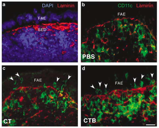
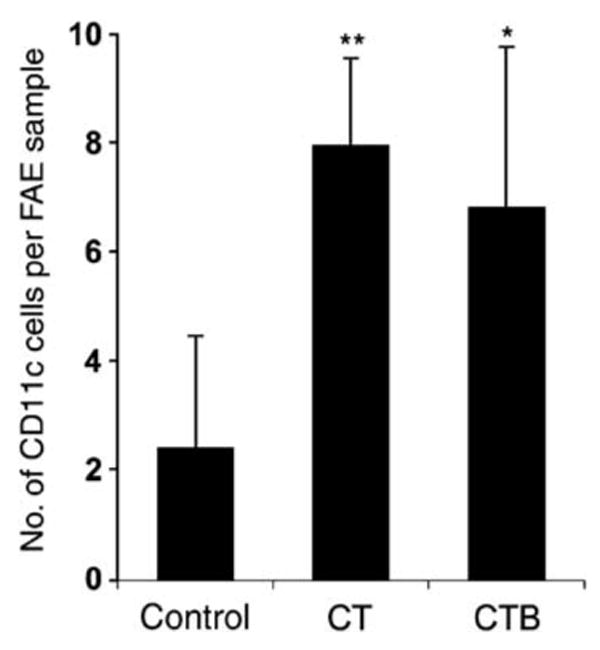
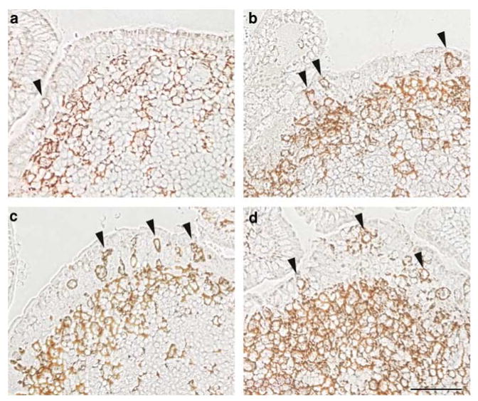
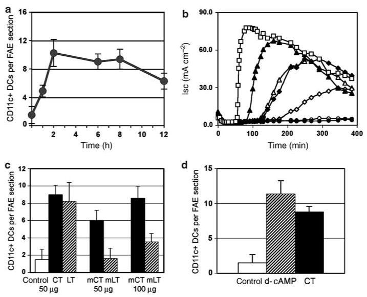
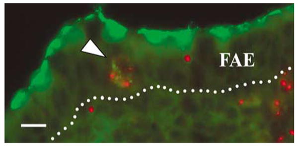
The follicle-associated epithelium (FAE) of Peyer's patches (PPs) transports antigens and microorganisms into mucosal lymphoid tissues where they are captured by subepithelial dendritic cells (DCs). Feeding of cholera toxin (CT) induced migration of subepithelial DCs to interfollicular T-cell areas within 24 h. This study investigated short-term effects of CT, Escherichia coli heat-labile toxin, and non-toxic derivatives on DC migration. CT or CTB injected into ligated intestinal loops induced significant increase in CD11c+ DCs within the FAE within 90 min. In mice fed CT intragastrically, DC numbers in the FAE increased by 1 h, were maximal by 2 h, declined between 8 and 12 h, and were reversed by 24 h. Feeding of native LT, recombinant CTB, dibutyryl cyclic AMP, and to a lesser extent mutated CT(E29H) or mutated LT(R192G) had the same effect. Thus, both A and B subunits of enterotoxins, presumably acting through distinct signaling pathways, may promote capture of incoming antigens and pathogens by PP DCs.


Conflict of interest statement

Disclosure: The authors declared no conflict of interest.

Figures

Figure 1
Figure 1
CT and CTB injected into ligated ileal loops induce migration of DCs into the FAE. (a) Control PP tissue section stained with DAPI (blue) and anti-laminin (red) to visualize the boundary between the SED region and the FAE. (b–d) Sections of PP domes stained for CD11c (green) and laminin (red), taken from ligated ileal loops injected with (b) PBS, (c) CT, or (d) CTB 90 min earlier. Increased numbers of DCs (arrowheads) are present in the FAE following injection of either CT or CTB. The images shown are representative of sections from four BALB/c mice per group. CT, cholera toxin; DAPI, 4,6-diamidino-2-phenylindole; DCs, dendritic cells; FAE, follicle-associated epithelium; PBS, phosphate-buffered saline; PPs, Peyer's patches; SED, subepithelial dome. Bar = 10μm.
Figure 2
Figure 2
Quantitation of CD11c + DCs in the FAE of BALB/c mice (n=4) 90 min after intraluminal injection of CT, CTB, or PBS (control). Values are expressed as mean±s.d. of intraepithelial DCs per standard area as defined in Methods (*P<0.05; **P<0.001, treated vs. control). CT, cholera toxin; DCs, dendritic cells; FAE, follicle-associated epithelium; PBS, phosphate-buffered saline.
Figure 3
Figure 3
CT, LT, and rCTB fed perorally induce migration of CD11c + DCs into the FAE of PP. Groups of mice (n=4) were fed (a) PBS, or 50 μg of either (b) CT, (c) LT, or (d) rCTB, and distal PPs were collected 6 h later. Sections were treated with biotinylated anti-CD11c antibodies followed by avidin–HRP to visualize DCs within the FAE (arrowheads) and in the subepithelial dome region. CD11c + DCs in the FAE and in the SED region were increased in mice fed with CT, LT, or rCTB. CT, cholera toxin; DCs, dendritic cells; FAE, follicle-associated epithelium; HRP, horseradish peroxidase; PBS, phosphate-buffered saline; PPs, Peyer's patches; rCTB, recombinant CTB; SED, subepithelial dome. Bar = 50μm.
Figure 4
Figure 4
Analysis of DC migration effects induced by feeding of enterotoxins and derivatives. (a) Time course of DC migration into the FAE of PP after feeding native CT. Groups of mice (n=4) were fed 50 μg of CT and killed at 1, 2, 6, 8, or 12h post-feeding, and sections from four distal PPs from each mouse were stained with anti-CD11c antibodies. Mean numbers (±s.d.) of CD11c + cells per FAE section for each group are shown. (b) Analysis of ADP-ribosyltransferase activity in the native and mutant enterotoxin preparations fed perorally, measured as elevation of short-circuit current (Isc) across confluent monolayers of T84 intestinal epithelial cells grown on Transwell filters. Results of a representative experiment of three are shown. The curves reflect elevation of intracellular cAMP produced by 10 nm of native CT (filled triangles), native LT (filled diamonds), mutant CT(E29H) (open triangles), mutant LT(R192G) (open diamonds), or rLTB (filled circles). A 1 mm solution of d-cAMP (open squares) served as a positive control and buffer alone (open circles) served as a negative control. (c) Numbers of DCs per FAE section in groups of mice (n=4) fed either PBS (cont) or 50 μg of native CT, native LT, mutant CT(E29H) (mCT), mutant LT(R192G) (mLT), or 100 μg of mutant toxins. (Bars represent mean±s.d.) After 50 μg of LT(R192G), numbers of DCs in the FAE were unchanged relative to controls. In mice fed 50 μg of CT(E29H), the average number of DCs per FAE section was increased but was significantly lower than that in mice treated with 50 μg of native CT (P<0.0001). Hundred micrograms of CT(E29H) had an effect comparable to that of 50 μg native CT or LT (P=0.3367). Hundred micrograms of LT(R192G) had a significantly smaller effect than the same dose of CT(E29H) (P<0.0001). (d) Injection of d-cAMP into ligated ileal loops mimics the effect of native CT on DC movement into the FAE. Mean numbers (±s.d.) of CD11c + DCs per FAE of PP from groups of mice (n=4) inoculated via ligated loops with 1 mM d-cAMP (cross-hatched bar), 50 μg ml−1 CT (filled bar), or saline (open bar) are shown. CT, cholera toxin; DAPI, 4,6-diamidino-2-phenylindole; d-cAMP, dibutyryl cyclic AMP; DCs, dendritic cells; FAE, follicle-associated epithelium; PBS, phosphate-buffered saline; PPs, Peyer's patches.
Figure 5
Figure 5
DCs loaded with fluorescent microparticles take up live reovirus in the FAE. Mice were fed red fluorescent microparticles, and 24 h later, ligated ileal loops were inoculated with live reovirus. PPs were collected and fixed 1 h later, and sections were stained with rabbit anti-reovirus serum followed by secondary FITC-conjugated antibodies. Reovirus (green) is concentrated at the apical poles of FAE cells. Some reovirus is colocalized with red fluorescent microparticles in a cell within the FAE (arrowhead). The dotted line indicates the basal surface of the FAE. DCs, dendritic cells; FAE, follicle-associated epithelium; FITC, fluorescein isothiocyanate; PPs, Peyer's patches. Bar = 10 μm.

References

    1. Strober W, McGhee JR. Inductive and effector tissues and cells of the mucosal immune system. In: Mestecky J, Lamm M, McGhee J, Bienenstock J, Mayer L, Strober W, editors. Mucosal Immunology. Academic Press; New York: 2005. pp. 371–374.
    1. Neutra MR, Mantis NJ, Kraehenbuhl JP. Collaboration of epithelial cells with organized mucosal lymphoid tissues. Nat Immunol. 2001;2:1004–1009. - PubMed
    1. Kelsall BL, Strober W. Distinct populations of dendritic cells are present in the subepithelial dome and T cell regions of murine Peyer's patches. J Exp Med. 1996;183:237–247. - PMC - PubMed
    1. Iwasaki A, Kelsall BL. Localization of distinct Peyer's patch dendritic cell subsets and their recruitment by chemokines macrophage inflammatory protein (MIP)-3alpha, MIP-3beta, and secondary lymphoid organ chemokine. J Exp Med. 2000;191:1381–1394. - PMC - PubMed
    1. Cook DN, Prosser DM, Forster R, Zhang J, Kuklin NA, Abbondanzo SJ, et al. CCR6 mediates dendritic cell localization, lymphocyte homeostasis, and immune responses in mucosal tissue. Immunity. 2000;12:495–503. - PubMed

Publication types

MeSH terms