. 2008 Jan;1(1):68-77.
doi: 10.1038/mi.2007.2.

Distal IgA immunity can be sustained by alphaEbeta7+ B cells in L-selectin-/- mice following oral immunization

Affiliations

Distal IgA immunity can be sustained by alphaEbeta7+ B cells in L-selectin-/- mice following oral immunization

D W Pascual et al. Mucosal Immunol. 2008 Jan.

Abstract

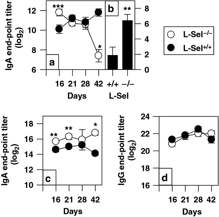
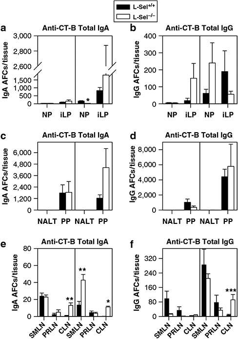
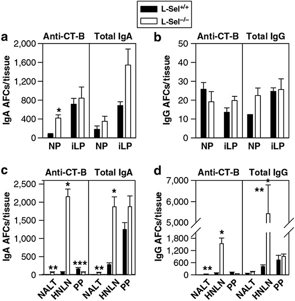
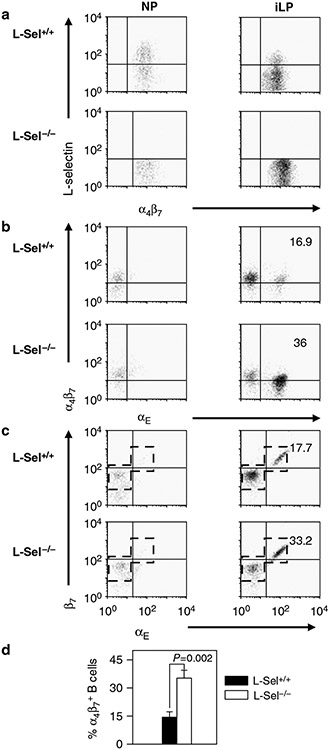
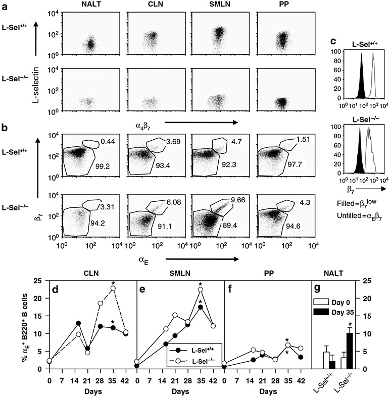
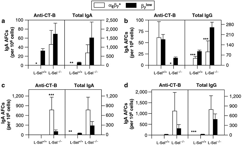
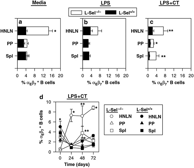
Understanding the role of homing receptors could aid vaccine strategies for developing distal mucosal immunity. Infection studies have revealed that immune intestinal B cells use alpha(4)beta(7) homing receptors, but their role in subsequent oral immunization with soluble antigens is unknown. To assess the influence of L-selectin and alpha(4)beta(7) on distal B cells following oral cholera toxin (CT) immunization, L-selectin-deficient (L-Sel(-/-)) IgA anti-CT-B-specific B cells were enhanced 30-, 9.2-, and 3.5-fold in head and neck lymph nodes (HNLNs), nasal-associated lymphoid tissue, and nasal passages (NPs), respectively, vs. L-Sel(+/+) mice. Cell-sorted intestinal and NP IgA antibody-forming cells (AFCs) were mostly alpha(4)beta(7)(+), unlike HNLN L-Sel(-/-) IgA and IgG anti-CT-B AFCs that were alpha(E)beta(7)(+), contrasting with L-Sel(+/+) HNLN IgA AFCs that were mostly alpha(4)beta(7)(+). In vitro studies revealed that L-Sel(-/-) HNLN B cells preferentially expressed alpha(E) following polyclonal stimulation. These studies show that HNLN B cells express alpha(E)beta(7) in the absence of L-selectin to sustain distal IgA responses.


Conflict of interest statement

DISCLOSURE

The authors declared no conflict of interest.

Figures

Figure 1
Figure 1
Rapid onset mucosal IgA in L-Sel−/− mice. Depicted are (a) fecal IgA and serum (c) IgA and (d) IgG anti-CT-B titers following oral immunization of L-Sel−/− mice with CT. L-Sel−/− and L-Sel+/+ mice were orally immunized on days 0, 7, and 14. Serum and mucosal secretions were collected and assessed for immune Abs by CT-B ELISA. Although L-Sel−/− fecal IgA anti-CT-B Abs decreased at 42 days, (b) S-IgA in nasal washes from L-Sel−/− mice remained elevated. Results up to 42 days after primary immunization are depicted as the mean of 10 mice ±s.e.m., and statistical differences between L-Sel−/− and L-Sel+/+ mice were determined: *P < 0.001; **P = 0.006; ***P = 0.021. Ab, antibody; CT, cholera toxin; ELISA, enzyme-linked immunosorbent assay; Ig, immunoglobulin; L-Sel, L-selectin.
Figure 2
Figure 2
Oral immunization of L-Sel−/− mice with CT results in increased IgA AFC responses in CLN 16 days post-primary immunization. L-Sel−/− and L-Sel+/+ mice were orally immunized, as previously described, and on day 16, total lymphocytes from (a, b) NP and small iLP, (c, d) NALT and PP, and (e, f) SMLN, PRLN, and CLN were collected and assayed for anti-CT-B and total (a, c, e) IgA and (b, d, f) IgG AFC responses. CT-B-specific and total IgA AFC responses were slightly enhanced in L-Sel−/− CLN as well as total IgG AFCs. The number of IgA and IgG CT-B-specific AFCs in the other L-Sel−/− HNLN, NALT, PP, NP, and iLP were not significantly different from L-Sel+/+ mice. Results depict the mean of three experiments ±s.e.m. per tissue, and statistical differences between L-Sel−/− and L-Sel+/+ mice were determined: *P < 0.001; **P ⩽ 0.003; ***P = 0.031. AFC, antibody-forming cell; CLN, cervical lymph node; CT, cholera toxin; HNLN, head and neck lymph node; Ig, immunoglobulin; iLP, intestinal lamina propria; L-Sel, L-selectin; NALT, nasal-associated lymphoid tissue; NP, nasal passage; PP, Peyer’s patch; PRLN, parotid gland lymph node; SMLN, submaxillary gland lymph node.
Figure 3
Figure 3
Oral immunization of L-Sel−/− mice with CT results in enhanced IgA AFC responses in NP 42 days post-primary immunization and supported by increases in NALT and HNLN IgA AFCs. L-Sel−/− and L-Sel+/+ mice were orally immunized, as previously described, and on day 42, total lymphocytes from (a, b) NP and iLP and (c, d) NALT, HNLN, and PP were collected and assayed for anti-CT-B and total (a, c) IgA and (b, d) IgG AFC responses. Each L-Sel−/− HNLN was combined and assessed for anti-CT-B activity. CT-B-specific IgA AFC responses were elevated in L-Sel−/− mice when compared to L-Sel+/+ mice. IgA and IgG iLP CT-B-specific AFC responses were not significantly different between L-Sel+/+ and L-Sel−/− mice; however, dramatic increases in L-Sel−/− NALT and HNLN CT-B-specific IgA and IgG AFC responses were observed when compared to L-Sel+/+ mice. Total IgA and IgG AFCs in L-Sel−/− HNLN and total IgG AFCs in L-Sel−/− NALT were also elevated when compared to L-Sel+/+ mice. Depicted are the mean AFC responses of three experiments ±s.e.m. per tissue, and statistical differences between L-Sel−/− and L-Sel+/+ mice were determined: *P < 0.001; **P ⩽ 0.014; ***P = 0.025. AFC, antibody-forming cell; CT, cholera toxin; HNLN, head and neck lymph node; Ig, immunoglobulin; iLP, intestinal lamina propria; L-Sel, L-selectin; NALT, nasal-associated lymphoid tissue; NP, nasal passage; PP, Peyer’s patch; PRLN, parotid gland lymph node; SMLN, submaxillary gland lymph node.
Figure 4
Figure 4
Increased presence of αEβ7+ iLP B cells in the L-Sel−/− mice 6 weeks post-primary oral immunization with CT. L-Sel+/+ and L-Sel−/− NP and iLP B cells were analyzed for expression of (a) L-Sel and α4β7, (b) α4β7 and αE, and (c) β7 and αE. αEβ7+ B cells remained as a minor subset in the NP, but in the L-Sel−/− iLP they showed (d) ~2.4-fold greater percentage than that in L-Sel+/+ iLP. (c) Boxed histographs represent the populations sorted for B-cell ELISPOT in Figure 5. Data are representative of three experiments except in d, which shows the mean±s.e.m. of three experiments. CT, cholera toxin; ELISPOT, enzyme-linked immunosorbent spot; iLP, intestinal lamina propria; L-Sel, L-selectin; NP, nasal passage.
Figure 5
Figure 5
Effector CT-B-specific B cells were mostly L-Sellow7low in the iLP and NP at 42 days post-primary immunization. Cell-sorting experiments were conducted sorting (a, b) iLP and (c, d) NP lymphocytes for β7 vs. β7high and αE expression and assessed by CT-B-specific (left panel) and total (right panel) (a, c) IgA and (b, d) IgG ELISPOT. No β7high B cells were found in either L-Sel+/+ or L-Sel−/− NP; all the iLP β7high B cells were αEβ7+, not α4β7high, and the β7low B cells were all L-Sellow7low. The majority of the CT-B-specific iLP IgA AFCs were β7low. For both L-Sel+/+ and L-Sel−/− mice, the L-Sellow7low B-cell subset contained all of the NP IgA anti-CT-B activity, and for L-Sel−/− mice, αEβ7+ B cells contained the IgG anti-CT-B and the total IgG AFCs. Results depict the mean of three experiments ±s.e.m., and statistical differences between β7low and αEβ7+ B cells were determined: *P ⩽ 0.002; **P ⩽ 0.008; ***P < 0.026; ****P < 0.05. AFC, antibody-forming cell; CT, cholera toxin; ELISPOT, enzyme-linked immunosorbent spot; Ig, immunoglobulin; iLP, intestinal lamina propria; L-Sel, L-selectin; NALT, nasal-associated lymphoid tissue; NP, nasal passage.
Figure 6
Figure 6
αEβ7+ B cells appear late in mucosal inductive tissues after primary oral immunization with CT in both L-Sel+/+ and L-Sel−/− mice. L-Sel+/+ and L-Sel−/− NALT, CLN, SMLN, and PP B cells were analyzed for expression of (a) L-Sel and α4β7 and (b) β7 and αE at 6 weeks post-primary immunization. Expression for αEβ7 was evident in each of the mucosal inductive tissues examined. (c) Cell-sorting profiles show that the sorted β7low vs. αEβ7 B cells were greater than 95% pure. (d–g) A kinetic analysis was performed to determine when these αEβ7+ B cells are induced in the (d) CLN, (e) SMLN, (f) PP, and (g) NALT. Less than 5% αEβ7+ B cells are present in naive mucosal inductive tissues, but these increase with time, peaking at day 35 post-primary immunization. Each time point represents pooled tissues from five individual mice, and differences between days 0 and 35 were evaluated: *P < 0.001. CLN, cervical lymph node; CT, cholera toxin; ELISPOT, enzyme-linked immunosorbent spot; Ig, immunoglobulin; iLP, intestinal lamina propria; L-Sel, L-selectin; NALT, nasal-associated lymphoid tissue; NP, nasal passage; PP, Peyer’s patch; SMLN, submaxillary gland lymph node.
Figure 7
Figure 7
The increased numbers of B cells in the L-Sel−/− HNLN at 42 days post-primary immunization are attributed to increased functional activity by αEβ7+ B cells exhibiting both IgA and IgG anti-CT-B activity. Cell-sorting experiments were conducted on (a, b) PP and (c, d) HNLN lymphocytes for β7 vs. β7high and αE expression and assessed by CT-B-specific (left panel) and total (right panel) (a, c) IgA and (b, d) IgG ELISPOT. (a, b) For PP CT-B-specific responses, all the L-Sel+/+ IgA AFCs were α4β7low, and IgG AFCs were equally divided between α4β7low and αEβ7+ subsets. The L-Sel−/− PP IgA AFCs were equally divided between both subsets, and no αEβ7+ IgG anti-CT-B AFCs were detected. (c, d) For the HNLN CT-B-specific responses, very few to no IgA or IgG AFCs were detected in L-Sel+/+ mice. In contrast, the majority of the IgA AFCs were αEβ7+, and the IgG AFC responses were composed of either αEβ7+ or α4β7low subset. Thus, sustained and elevated IgA anti-CT-B responses are attributed to the αEβ7+ Ag-reactive B cells found late in L-Sel−/− HNLN following oral immunization with CT. The results depict the mean of three experiments ±s.e.m., and statistical differences between β7low and αEβ7+ B cells were determined: *P ⩽ 0.008; **P = 0.02; ***P ⩽ 0.048. AFC, antibody-forming cell; Ag, antigen; CT, cholera toxin; ELISPOT, enzyme-linked immunosorbent spot; HNLN, head and neck lymph node; Ig, immunoglobulin; L-Sel, L-selectin; PP, Peyer’s patch.
Figure 8
Figure 8
L-Sel−/− HNLN shows increased expression of αEβ7+ B cells. Total HNLN, PP, and Spl lymphocytes were cultured (a) without activation or activated with (b) LPS or (c) LPS plus CT for 48 h. B220+ TCR-β lymphocytes were then analyzed for αE and β7 expression. Increased percentages of αEβ7+ B cells were observed with the (a) HNLN lymphocytes left unstimulated and (c) HNLN, PP, and Spl lymphocytes stimulated with LPS plus CT. (d) A kinetic analysis was performed and showed that L-Sel−/− HNLN αEβ7+ B cells showed preferential enhancement at 24, 48, and 72 h with LPS plus CT treatment. Depicted data are the means of six individual mice ±s.e.m., and statistical differences between L-Sel−/− and L-Sel+/+ mice were determined: *P ⩽ 0.001; **P ⩽ 0.015. CT, cholera toxin; HNLN, head and neck lymph node; LPS, lipopolysaccharide; L-Sel, L-selectin; PP, Peyer’s patch; Spl, splenic; TCR-β, T-cell receptor-β.

Similar articles

Cited by

References

    1. Rudzik R, Clancy RL, Perey DY, Day RP & Bienenstock J Repopulation with IgA-containing cells of bronchial and intestinal lamina propria after transfer of homologous Peyer’s patch and bronchial lymphocytes. J. Immunol 114, 1599–1604 (1975). - PubMed
    1. Mestecky J, McGhee JR, Michalek SM, Arnold RR, Crago SS & Babb JL Concept of the local and common mucosal immune response. Adv. Exp. Med. Biol 107, 185–192 (1978). - PubMed
    1. McDermott MR & Bienenstock J Evidence for a common mucosal immunologic system. I. Migration of B immunoblasts into intestinal, respiratory, and genital tissues. J. Immunol 122, 1892–1898 (1979). - PubMed
    1. Quiding-Jarbrink M, Nordstrom L, Granstrom G, Kilander A, Jertbom M, Butcher EC et al. Differential expression of tissue-specific adhesion molecules on human circulating antibody-forming cells after systemic, enteric, and nasal immunizations. A molecular basis for the compartmentalization of effector B cell responses. J. Clin. Invest 99, 1281–1286 (1997). - PMC - PubMed
    1. Kantele A, Hakkinen M, Moldoveanu Z, Lu A, Savilahti E, Alvarez RD et al. Differences in immune responses induced by oral and rectal immunizations with Salmonella typhi Ty21a: evidence for compartmentalization within the common mucosal immune system in humans. Infect. Immun 66, 5630–5635 (1998). - PMC - PubMed

Publication types

MeSH terms

LinkOut - more resources