

Gene-centric association mapping of chromosome 3p implicates MST1 in IBD pathogenesis
- PMID: 19079170
- PMCID: PMC4550306
- DOI: 10.1038/mi.2007.15
Gene-centric association mapping of chromosome 3p implicates MST1 in IBD pathogenesis
Abstract
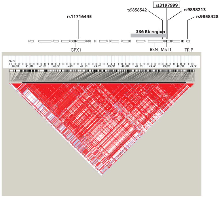
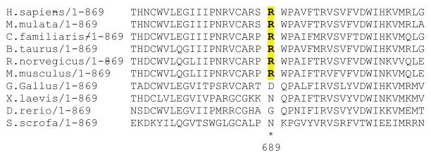
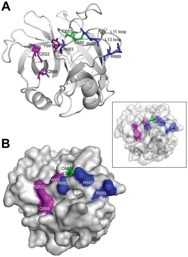
Association mapping and candidate gene studies within inflammatory bowel diseases (IBD) linkage regions, as well as genome-wide association studies in Crohn's disease (CD) have led to the discovery of multiple risk genes, but these explain only a fraction of the genetic susceptibility observed in IBD. We have thus been pursuing a region on chromosome 3p21-22 showing linkage to CD and ulcerative colitis (UC) using a gene-centric association mapping approach. We identified 12 functional candidate genes by searching for literature cocitations with relevant keywords and for gene expression patterns consistent with immune/intestinal function. We then performed an association study composed of a screening phase, where tagging single nucleotide polymorphisms (SNPs) were evaluated in 1,020 IBD patients, and an independent replication phase in 745 IBD patients. These analyses identified and replicated significant association with IBD for four SNPs within a 1.2 Mb linkage disequilibrium region. We then identified a non-synonymous coding variant (rs3197999, R689C) in the macrophage-stimulating 1 (MST1) gene (P-value 3.62 x 10(-6)) that accounts for the association signal, and shows association with both CD and UC. MST1 encodes macrophage-stimulating protein (MSP), a protein regulating the innate immune responses to bacterial ligands. R689C is predicted to interfere with MSP binding to its receptor, suggesting a role for this gene in the pathogenesis of IBD.
Figures
References
-
- Binder V. Genetic epidemiology in inflammatory bowel disease. Dig Dis. 1998 Nov-Dec;16(6):351–5. - PubMed
-
- Orholm M, Munkholm P, Langholz E, Nielsen OH, Sorensen TI, Binder V. Familial occurrence of inflammatory bowel disease. N Engl J Med. 1991 Jan 10;324(2):84–8. - PubMed
-
- Satsangi J, Parkes M, Louis E, Hashimoto L, Kato N, Welsh K, et al. Two stage genome-wide search in inflammatory bowel disease provides evidence for susceptibility loci on chromosomes 3, 7 and 12. Nat Genet. 1996 Oct;14(2):199–202. - PubMed
Publication types
MeSH terms
Substances
Grants and funding
- AI062773/AI/NIAID NIH HHS/United States
- P30 DK040561/DK/NIDDK NIH HHS/United States
- DK62413/DK/NIDDK NIH HHS/United States
- U01 DK062413/DK/NIDDK NIH HHS/United States
- U24 DK062429/DK/NIDDK NIH HHS/United States
- DK064869/DK/NIDDK NIH HHS/United States
- DK62431/DK/NIDDK NIH HHS/United States
- U01 DK062432/DK/NIDDK NIH HHS/United States
- R01 DK064869/DK/NIDDK NIH HHS/United States
- U01 DK062429/DK/NIDDK NIH HHS/United States
- U01 DK062422/DK/NIDDK NIH HHS/United States
- DK62422/DK/NIDDK NIH HHS/United States
- R01 AI062773/AI/NIAID NIH HHS/United States
- U01 DK062423/DK/NIDDK NIH HHS/United States
- DK62432/DK/NIDDK NIH HHS/United States
- DK62423/DK/NIDDK NIH HHS/United States
- DK62429/DK/NIDDK NIH HHS/United States
- DK62420/DK/NIDDK NIH HHS/United States
- U01 DK062420/DK/NIDDK NIH HHS/United States
- U01 DK062431/DK/NIDDK NIH HHS/United States
LinkOut - more resources
Full Text Sources
Other Literature Sources
Medical
Molecular Biology Databases
Research Materials
Miscellaneous
