Review
. 2008 May;1(3):183-97.
doi: 10.1038/mi.2008.5. Epub 2008 Mar 5.

Mucins in the mucosal barrier to infection

Affiliations
Review

Mucins in the mucosal barrier to infection

S K Linden et al. Mucosal Immunol. 2008 May.

Abstract

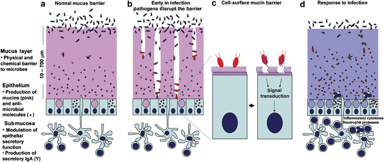
The mucosal tissues of the gastrointestinal, respiratory, reproductive, and urinary tracts, and the surface of the eye present an enormous surface area to the exterior environment. All of these tissues are covered with resident microbial flora, which vary considerably in composition and complexity. Mucosal tissues represent the site of infection or route of access for the majority of viruses, bacteria, yeast, protozoa, and multicellular parasites that cause human disease. Mucin glycoproteins are secreted in large quantities by mucosal epithelia, and cell surface mucins are a prominent feature of the apical glycocalyx of all mucosal epithelia. In this review, we highlight the central role played by mucins in accommodating the resident commensal flora and limiting infectious disease, interplay between underlying innate and adaptive immunity and mucins, and the strategies used by successful mucosal pathogens to subvert or avoid the mucin barrier, with a particular focus on bacteria.


Figures

Figure 1
Figure 1
Diagrammatic representation of mucins in the mucosal barrier to infection. (a) The normal mucosa is covered with a continuously replenished thick mucus layer retaining host-defensive molecules. Commensal and environmental microbes may live in the outer mucus layer but the layer ensures that contact of microbes with epithelial cells is rare. (b) Early in infection, many pathogens actively disrupt the mucus layer and thereby gain access to the epithelial cell surface. In addition, this alters the environment for commensal and environmental microbes and opportunistic pathogenesis may occur. (c) Pathogens that break the secreted mucus barrier reach the apical membrane surface, which is decorated with a dense network of large cell-surface mucins. Pathogens bind the cell-surface mucins via lectin interactions and the mucin extracellular domains are shed as releasable decoy molecules. Consequent to contact with microbes and shedding of the extracellular domain, signal transduction by the cytoplasmic domains of the cell-surface mucins modulates cellular response to the presence of microbes. (d) In response to infection, there are alterations in mucins that are driven directly by epithelial cells and in response to signals from underlying innate and adaptive immunity. These alterations include goblet cell hyperplasia and increased mucin secretion and altered mucin glycosylation (depicted by the color change) affecting microbial adhesion and the ability of microbes to degrade mucus. These changes in mucins work in concert with other arms of immunity to clear the infection. PowerPoint slide

Similar articles

Cited by

References

    1. Kagnoff MF, Eckmann L. Epithelial cells as sensors for microbial infection. J. Clin. Invest. 1997;100,:6–10. doi: 10.1172/JCI119522. - DOI - PMC - PubMed
    1. Lu J, Teh C, Kishore U, Reid KB. Collectins and ficolins: sugar pattern recognition molecules of the mammalian innate immune system. Biochim. Biophys. Acta. 2002;1572,:387–400. doi: 10.1016/S0304-4165(02)00320-3. - DOI - PubMed
    1. Raj PA, Dentino AR. Current status of defensins and their role in innate and adaptive immunity. FEMS Microbiol. Lett. 2002;206,:9–18. doi: 10.1111/j.1574-6968.2002.tb10979.x. - DOI - PubMed
    1. Prydal JI, Muir MG, Dilly PN. Comparison of tear film thickness in three species determined by the glass fibre method and confocal microscopy. Eye. 1993;7,:472–475. doi: 10.1038/eye.1993.96. - DOI - PubMed
    1. Mercer RR ML R, Crapo JD. Mucous lining layers in human and rat airways. Ann. Rev. Resp. Dis. 1992;145,:355. doi: 10.1164/ajrccm/145.2_Pt_1.355. - DOI

LinkOut - more resources