. 2008 May;1(3):229-38.
doi: 10.1038/mi.2008.1. Epub 2008 Mar 5.

TREM-2 binds to lipooligosaccharides of Neisseria gonorrhoeae and is expressed on reproductive tract epithelial cells

Affiliations

TREM-2 binds to lipooligosaccharides of Neisseria gonorrhoeae and is expressed on reproductive tract epithelial cells

D N Quan et al. Mucosal Immunol. 2008 May.

Abstract

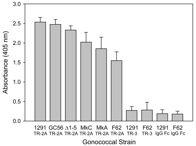
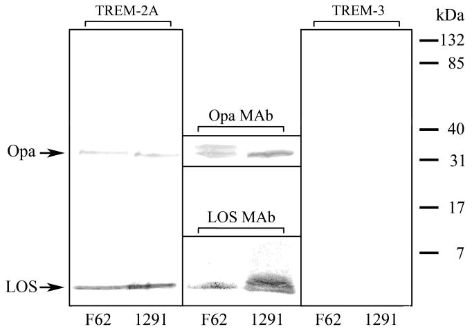
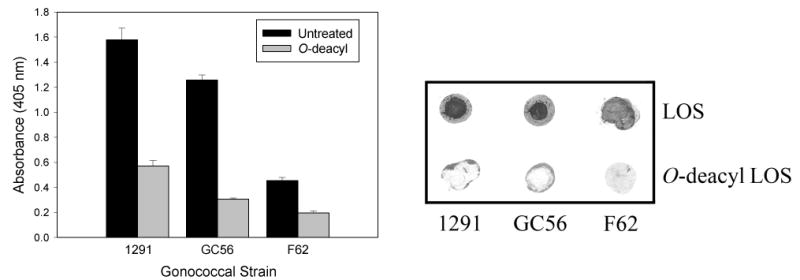
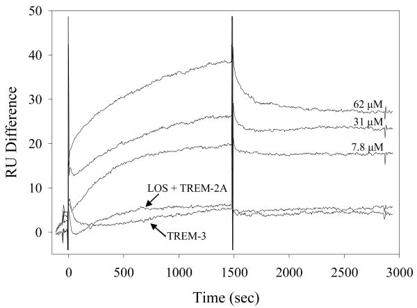
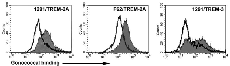
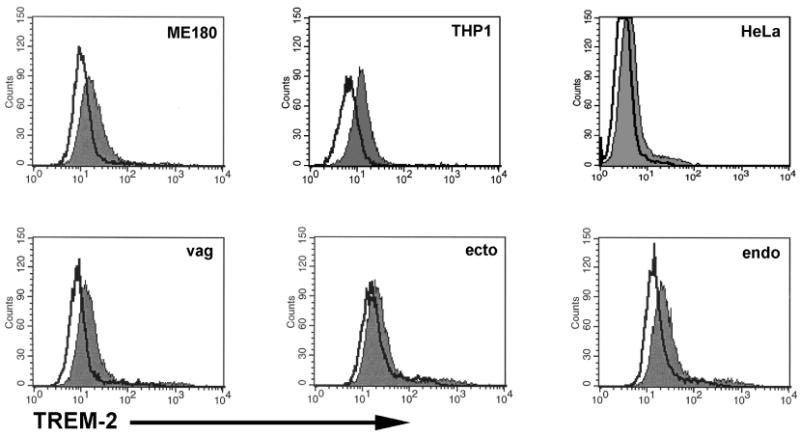
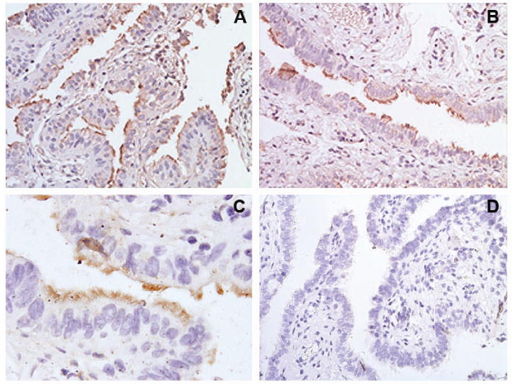
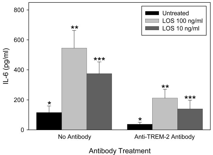
Triggering receptor expressed on myeloid cells-2 (TREM-2) is an innate immune receptor that initiates cellular activation upon ligation. In this study, we examined the interaction of TREM-2 with Neisseria gonorrhoeae using murine TREM-2A, as it has been reported to recognize bacterial ligands. Using a whole-bacteria enzyme-linked immunosorbent assay (ELISA), TREM-2A bound to all six strains in variable degrees. Far-western blots of gonococcal outer membranes revealed TREM-2A binding to lipooligosaccharide (LOS) and opacity (Opa) protein, with predominant binding to LOS. Binding of TREM-2A to LOS was confirmed by ELISA and surface plasmon resonance. O-deacylation of the lipid A significantly reduced binding. Flow cytometry and reporter cell assays showed that gonococci bound to TREM-2A-transfected cells and induced transmembrane signaling. In humans, TREM-2 was constitutively expressed by genitourinary and fallopian tube epithelial cells, both of which are primary targets of gonococcal invasion. Ligation of TREM-2 by LOS induced interleukin-6 production in HeLa cervical carcinoma cells. To our knowledge, this is the first report of the expression of human TREM-2 by cells deriving from a non-myeloid lineage. We conclude that gonococci can interact with TREM-2 receptors through binding to LOS and Opa protein and initiate cell signaling and cytokine production.


Figures

Figure 1
Figure 1
TREM-2A binds to various strains of N. gonorrhoeae. Whole-cell ELISA analyses were performed by coating microtiter wells with gonococci and incubating the wells with TREM-2A (TR-2A), TREM-3 (TR-3), or IgG Fc fragments. The bars represent the mean of triplicate data points and the error bars are ±SD. The results are representative of four independent experiments.
Figure 2
Figure 2
TREM-2A binds gonococcal outer membrane components LOS and Opa protein as shown by far western blots. The SDS-PAGE-separated LOS and proteins from OMC purified from gonococcal strains F62 and 1291 were transferred to nitrocellulose and reacted with TREM-2A (left panel) or TREM-3 (right panel). Both strains bound TREM-2A to outer membrane components LOS and Opa protein, however >75% of binding was to LOS as determined by densitometry. No binding of TREM-3 to outer membrane components was detected. The identity of LOS was determined with the use of LOS MAb 1B2 (lower center panel), which binds to the lacto-N-neotetraose terminal structure of the LOS expressed by strains F62 and 1291, and that of Opa proteins was determined with the use of Opa MAb B33 (upper center panel), which reacts with all Opa protein variants. For the LOS, intensity differences in LOS MAb staining are the result of differences between the two strains in the amount of LOS expressed terminating in lacto-N-neotetraose as shown in Table 1. For the Opa proteins, F62 expressed two variants, only one of which bound TREM-2A. The results are representative of three independent experiments.
Figure 3
Figure 3
TREM-2A binds to intact gonococcal LOS but not to O-deacylated LOS. Left Panel: ELISA analyses of the binding of TREM-2A to intact and O-deacylated LOS. Wells were coated with LOS, blocked and reacted with TREM-2A. TREM-2A bound to the intact LOS of the strains tested but to variable degrees. No correlation was observed between the length of the OS α chains and TREM-2A binding based on the structures in Table 1. O-deacylated LOS bound substantially less TREM-2A than untreated LOS. Statistical comparisons: p < 0.001 for all three LOS comparing untreated versus O-deacylated. The bars represent the mean of triplicate data points and the error bars are ±SD. The data are representative of three independent experiments. Right Panel: Dot blot analyses of the binding of TREM-2A to LOS and O-deacylated LOS. LOS was spotted onto Hybond nitrocellulose membranes, which were blocked and incubated with TREM-2A. The significant decrease in binding of TREM-2A to O-deacylated LOS suggested a critical role for lipid A in the binding of TREM-2A to LOS. The data are representative of three independent experiments.
Figure 4
Figure 4
Surface plasmon resonance kinetic analyses of the binding of 1291 LOS to TREM-2A performed on a Biacore 3000 system using CM5 sensor chips. Increasing concentrations of LOS were injected at 10 μl/min into a flow cell containing TREM-2A-coated and control surfaces. For TREM-3, the highest concentration of LOS (62 μM) was injected over the TREM-3-coated and control surfaces. All binding signals were corrected for non-specific binding by subtracting the signal of the control flow cell. For the sensorgram labeled as LOS and TREM-2A, equimolar concentrations of LOS and soluble TREM-2A were incubated for 30 min prior to injection over the TREM-2A and control surfaces.
Figure 5
Figure 5
Gonococci bind to TREM-2A-transfected cells to a greater degree than to TREM-3-transfected or nontransfected cells. Gonococcal strains 1291 and F62 were fluorescently labeled and incubated with TREM-2A-expressing, TREM-3-expressing or nonexpressing BWZ cells. Flow cytometric analyses were performed to assess the degree of binding to each cell type. In the panels labeled TREM-2A, the filled histograms represent TREM-2A-expressing cells and the open histograms are nonexpressing BWZ cells. In the panel labeled TREM-3, the filled histogram is TREM-2A-expressing cells and the open histogram is TREM-3-expressing cells. The data are representative of three independent experiments.
Figure 6
Figure 6
Gonococci and purified LOS engage membrane-expressed TREM-2A and induce transmembrane signaling. BWZ reporter cell constructs expressing either TREM-2A (TR-2A) or TREM-3 (TR-3) were used to measure LacZ activity as an indicator of signal transduction following incubation of cells with gonococci or LOS. For stimulation with gonococci, reporter cells were incubated with a bacteria to cell ratio of either 100:1 or 1:1. For LOS, TREM-2A-expressing reporter cells were treated with LOS concentrations of either 100 μg/ml or 1 μg/ml. The LOS and all strains of gonococci induced LacZ activity but to variable degrees. Significantly greater signaling was induced in cells expressing TREM-2A than in those expressing TREM-3. Statistical comparisons: p < 0.05 for all comparisons of lacZ activity induced by gonococci and LOS in TREM-2A-expressing cells versus nontransfected cells; p < 0.001 for the comparisons of lacZ activity induced by 1291 and F62 in TREM-2A-expressing cells versus TREM-3-expressing cells. The bars represent the mean of triplicate data points and the error bars are ±SD. The results are representative of four independent experiments.
Figure 7
Figure 7
Human reproductive tract epithelial cells constitutively express TREM-2. Expression of TREM-2 by cultured human ectocervical, endocervical, vaginal, ME-180, and HeLa cells was detected by flow cytometric analyses. THP-1 cells known to express TREM-2 were stained as a positive control. Filled histograms represent cells stained with an anti-human TREM-2 antibody and open histograms represent cells stained with an irrelevant isotype matched antibody. The results are representative of four independent experiments.
Figure 8
Figure 8
Human fallopian tube epithelial cells constitutively express TREM-2. Photomicrographs of human fallopian tube organ cultures stained immunohistochemically for the expression of TREM-2. Panels A-C: Sections from three different female subjects stained for TREM-2 expression. Each section shows the localization of TREM-2 in the apical side of the brown-stained epithelium lining the lumen with little stromal cell staining for the protein. Staining was patchy in distribution with groups of stained cells located adjacent to groups of unstained cells, and expression was detected predominantly on ciliated epithelial cells; original magnification, ×200 (panels A and B), ×600 (panel C). Panel D: Negative control section incubated with non-immune goat IgG and the secondary detection antibody. No staining of any cell type was observed in control sections; original magnification, ×200.
Figure 9
Figure 9
Ligation of TREM-2 by LOS induces IL-6 production in HeLa cells. HeLa cells were treated with LOS from strain 1291 at concentrations of 10 and 100 ng/ml for 12 hours. Control wells were treated with culture media only. For some experiments, cells were treated with affinity-purified goat anti-human TREM-2 polyclonal IgG prior to addition of the LOS. Supernatants from the LOS-treated and untreated cells in the presence and absence of anti-human TREM-2 were assayed for IL-6 by ELISA. The bars represent the mean of quadruplicate data points and the error bars are ±SEM. The results are representative of three independent experiments. Statistical comparisons: *, p > 0.1; **, p < 0.05; ***, p < 0.05.

Similar articles

Cited by

References

    1. Centers for Disease Control and Prevention. Sexually Transmitted Disease Surveillance. U S Department of Health and Human Services, Public Health Service. 2005
    1. Paavonen J. Pelvic inflammatory disease. From diagnosis to prevention. Dermatol Clin. 1998;16:747–756. - PubMed
    1. Cohen MS, et al. Reduction of concentration of HIV-1 in semen after treatment of urethritis: implications for prevention of sexual transmission of HIV-1. Lancet. 1997;349:1868–1873. - PubMed
    1. Chesson HW, Pinkerton SD. Sexually transmitted diseases and the increased risk for HIV transmission: implications for cost-effectiveness analyses of sexually transmitted disease prevention interventions. J Acquir Immune Defic Syndr. 2000;24:48–56. - PubMed
    1. Mabey D. Interactions between HIV infection and other sexually transmitted diseases. Trop Med Int Health. 2000;5:A32–A36. - PubMed

Publication types

MeSH terms