. 2009 Feb 5;457(7230):736-40.
doi: 10.1038/nature07641. Epub 2008 Dec 14.

Messenger RNA targeting to endoplasmic reticulum stress signalling sites

Affiliations

Messenger RNA targeting to endoplasmic reticulum stress signalling sites

Tomás Aragón et al. Nature. .

Abstract

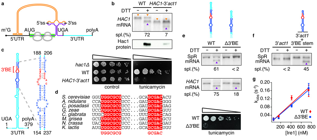
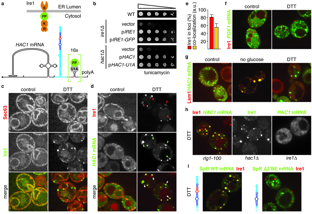
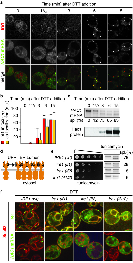
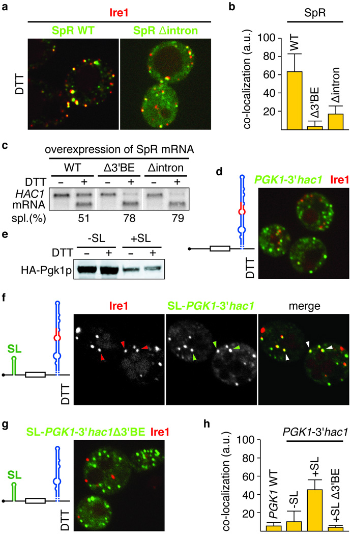
Deficiencies in the protein-folding capacity of the endoplasmic reticulum (ER) in all eukaryotic cells lead to ER stress and trigger the unfolded protein response (UPR). ER stress is sensed by Ire1, a transmembrane kinase/endoribonuclease, which initiates the non-conventional splicing of the messenger RNA encoding a key transcription activator, Hac1 in yeast or XBP1 in metazoans. In the absence of ER stress, ribosomes are stalled on unspliced HAC1 mRNA. The translational control is imposed by a base-pairing interaction between the HAC1 intron and the HAC1 5' untranslated region. After excision of the intron, transfer RNA ligase joins the severed exons, lifting the translational block and allowing synthesis of Hac1 from the spliced HAC1 mRNA to ensue. Hac1 in turn drives the UPR gene expression program comprising 7-8% of the yeast genome to counteract ER stress. Here we show that, on activation, Ire1 molecules cluster in the ER membrane into discrete foci of higher-order oligomers, to which unspliced HAC1 mRNA is recruited by means of a conserved bipartite targeting element contained in the 3' untranslated region. Disruption of either Ire1 clustering or HAC1 mRNA recruitment impairs UPR signalling. The HAC1 3' untranslated region element is sufficient to target other mRNAs to Ire1 foci, as long as their translation is repressed. Translational repression afforded by the intron fulfils this requirement for HAC1 mRNA. Recruitment of mRNA to signalling centres provides a new paradigm for the control of eukaryotic gene expression.


Figures

Figure 1
Figure 1. A conserved element in the 3’UTR of HAC1 mRNA is required for splicing in vivo, but not in vitro
a, Schematic of HAC1 mRNA. The Hac1 ORF is divided into two exons (purple). The intron (orange) base pairs with the 5′UTR (black), causing ribosome stalling (grey). Ire1 cleaves the intron at the indicated splice sites (5′ss & 3′ss). The green bar depicts where the GFP ORF replaces the HAC1 sequence in the splicing reporter. The 3′UTR is indicated in light blue. The 5′cap (m7G), start codon (AUG), stop codon (UGA) and polyadenylation signal (polyA) are indicated. b, e, f, Northern blot of HAC1 or SpR mRNA variants before or after ER stress induction with DTT (10 mM) for 45 min. Purple triangles denote spliced mRNAs; orange triangles denote unspliced mRNAs (only in b). Percent mRNA splicing (“Spl. (%)“) is indicated. Yeast strains harbor a genomic HAC1 copy with its own (WT) or ACT1’s 3′UTR sequence (HAC1-3′act1) (b, top), a genomic copy of SpR (e, top) or HAC1 (e, middle) bearing either the wild-type (WT) or the Δ3′BE mutant 3′UTR, as depicted, or a genomic copy of SpR with the 3’UTR of ACT1 with (3′act1+3′BE stem) or without (3′act1) an insertion of the 64 nucleotide element — shown in expanded view in (c) — as depicted (f). b, middle, Western blot of HA-tagged Hac1 protein from lysates from strains as in (b, top). b,e, Viability assay by 1:5 serial dilutions of hac1Δ or strains as in (b, top) or (e, middle) spotted onto solid media with or without 0.2 µg/ml of the ER stress inducer tunicamycin. Plates were photographed after 3 days growing at 30°C. c, Schematic of the HAC1 3′UTR stem-loop structure with the 3’BE (red) in a region (dark blue) that is shown in expanded view to the right; positional numbering from UGA stop codon. d, alignment of the 3′BE in HAC1 homologues. g, An in vitro intron excision reaction was performed as described with Ire1 concentrations: 50 nM, 150 nM, 400 nM, 730 nM of wild-type (red diamonds) or Δ3′BE (blue squares) HAC1 mRNA as substrates.
Figure 2
Figure 2. In response to ER stress HAC1 mRNA localizes to Ire1 foci in a 3’BE-dependent manner
a, Schematic of Ire1 and HAC1 mRNA imaging constructs: Ire1 has an ER-luminal stress sensing domain (S) and a kinase (K) and endonuclease domain (E) at its cytosolic face. GFP or mCherry (FP) was inserted between the transmembrane region and the kinase domain (up). b, Viability assay under ER stress conditions (0.2 µg/ml tunicamycin) of wild-type or ire1Δ yeast complemented with either empty vector, or centromeric plasmids bearing a wild-type (pIRE1), or the GFP-tagged imaging copy of Ire1 (pIRE1-GFP) (top) or hac1Δ yeast complemented with either empty plasmid, or a 2-micron plasmid bearing a wild-type (pHAC1) or the U1A-tagged imaging copy of HAC1 (pHAC1-U1A) (bottom). c,d, Localization of Sec63-mCherry and Ire1-GFP (c) or Ire1-mCherry and HAC1U1A mRNA decorated with U1A-GFP (d) before (left panels, control) and after (right panels, DTT) induction of ER stress. Arrowheads in d, lower panels, denote Ire1/HAC1 mRNA foci. e, Histogram depicting the percentage of Ire1 signal in foci (red bar) and the C.I. for HAC1U1A mRNA recruitment into Ire1 foci expressed in arbitrary units (“a.u.”, yellow bar); means +/− s.e.m., n = 9. f, Localization of Ire1-mCherry and PGK1U1A mRNA decorated with U1A-GFP under normal (left panel, control) and ER stress (right panel, DTT) conditions. g, Localization of Lsm1-mCherry and HAC1U1A mRNA without stress (left panel, control), after nutrient starvation for 10 min (middle panel, no glucose), or after induction of ER stress (right panel, DTT). h,i, Localization of Ire1-mCherry, Ire1-GFP, or HAC1U1A or SpR having 16 U1A hairpins as HAC1U1A (SpRU1A) either with or without the Δ3′BE deletion after induction of ER stress (DTT). c–i, ER stress was induced with 10 mM DTT for 45 min; imaging was performed in ire1Δ cells, complemented with Ire1 imaging constructs, except in (h) cell were hac1Δ or rlg1-100.
Figure 3
Figure 3. The HAC1 mRNA/Ire1 foci are functional UPR signaling centers
a, Localization of Ire1-mCherry and HAC1U1A mRNA decorated with U1A-GFP. b, Quantitation of the percentage of Ire1 signal in foci (red bars) and of the C.I. for HAC1U1A mRNA recruitment into Ire1 foci (yellow bars; means +/− s.e.m., n = 5). c, Northern blot of HAC1 mRNA (top) and Western blot of Hac1 protein (bottom). a–c, Samples were taken at indicated times after induction of ER stress with 10 mM DTT. d, Schematic of Ire1 oligomerization via interfaces 1 and 2. e, Viability assay under ER stress conditions (0.2 µg/ml tunicamycin) and Northern blot of HAC1 mRNA harvested from ire1Δ yeast complemented with wild-type Ire1 or interface mutants before or after treatment with 1 µg/ml tunicamycin for 1 h. f, Localization of Sec63-mCherry, Ire1-GFP, and HAC1U1A mRNA. Imaging was performed in ire1Δ yeast complemented with wild type or mutants of Ire1 that are defective in dimarization at interface 1 (if1), 2 (if2) or both (if1/2), either GFP-tagged (top) or untagged (bottom). ER stress was induced: 10 mM DTT 45 min. Separate channels are displayed in Supplementary Fig. S3.
Figure 4
Figure 4. Translational repression is a prerequisite for mRNA targeting to Ire1 foci
a,d,f–g, Localization of Ire1-mCherry, and U1A-GFP decorated mRNA of either the wild-type splicing reporter, as in Figure 2i (SpR WT), or an intron-less variant (SpR Δintron) (a), or of PGK1U1A, bearing either the wild-type (d,f) or mutant Δ3′BE (g) 3′UTR stem loop of HAC1 mRNA, in combination with (f,g) or without (d) a small stem loop (SL) that confers translational repression in its 5′UTR, as schematically depicted. b, C.I for mRNA recruitment of WT and Δintron SpR variants into Ire1 foci (means +/− s.e.m., n = 5); bar for the Δ3′BE mutant as depicted in Figure 2i is shown for comparison. c, Northern blot of HAC1 mRNA from yeast strains that over-expressed variants of the SpR, as indicated. e, Western blot of the variants of HA-tagged Pgk1 protein (HA-Pgk1p) bearing the 3′UTR from HAC1 with or without a 5′UTR SL a,c–g, ER stress was induced with 10 mM DTT for 45 min. h, C.I. for mRNA recruitment into Ire1 foci of PGK1U1A wild-type — see Figure 2f — or variants shown in d,f,g (means +/− s.e.m., n = 3–5).

Comment in

Similar articles

Cited by

References

    1. Bernales S, Papa FR, Walter P. Intracellular signaling by the unfolded protein response. Annu. Rev. Cell Dev. Biol. 2006;22:487–508. - PubMed
    1. Ron D, Walter P. Signal integration in the endoplasmic reticulum unfolded protein response. Nat. Rev. Mol. Cell Biol. 2007;8:519–529. - PubMed
    1. van Anken E, Braakman I. Endoplasmic reticulum stress and the making of a professional secretory cell. Crit. Rev. Biochem. Mol. Biol. 2005;40:269–283. - PubMed
    1. Rüegsegger U, Leber JH, Walter P. Block of HAC1 mRNA translation by long-range base pairing is released by cytoplasmic splicing upon induction of the unfolded protein response. Cell. 2001;107:103–114. - PubMed
    1. Sidrauski C, Cox JS, Walter P. tRNA ligase is required for regulated mRNA splicing in the unfolded protein response. Cell. 1996;87:405–413. - PubMed

Publication types

MeSH terms