. 2009 Jan;11(1):46-55.
doi: 10.1038/ncb1811. Epub 2008 Dec 14.

Secreted Frizzled-related protein 2 is a procollagen C proteinase enhancer with a role in fibrosis associated with myocardial infarction

Affiliations

Secreted Frizzled-related protein 2 is a procollagen C proteinase enhancer with a role in fibrosis associated with myocardial infarction

Koichi Kobayashi et al. Nat Cell Biol. 2009 Jan.

Abstract

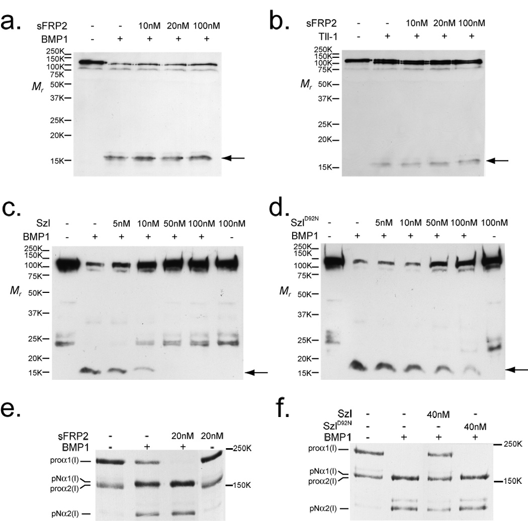
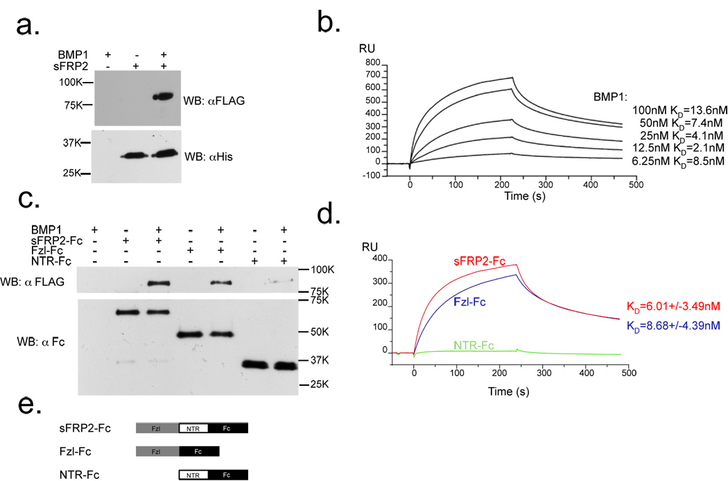
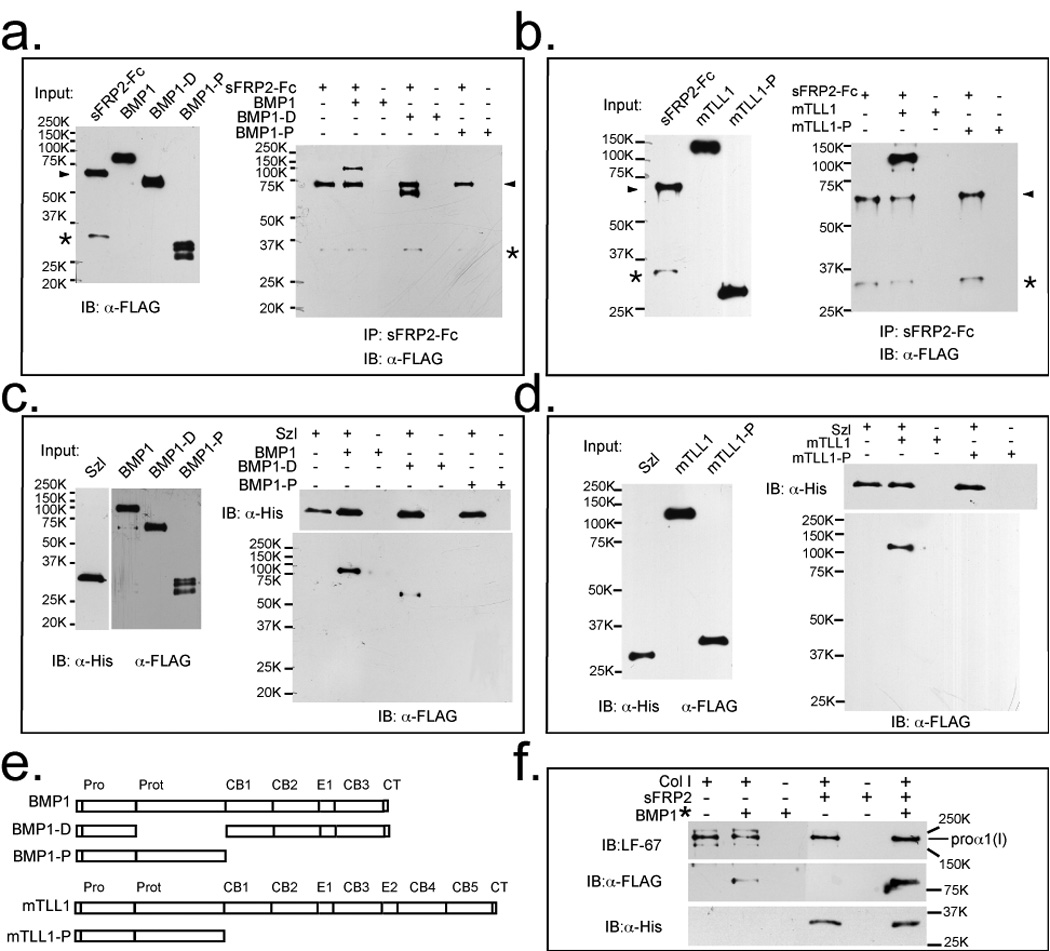
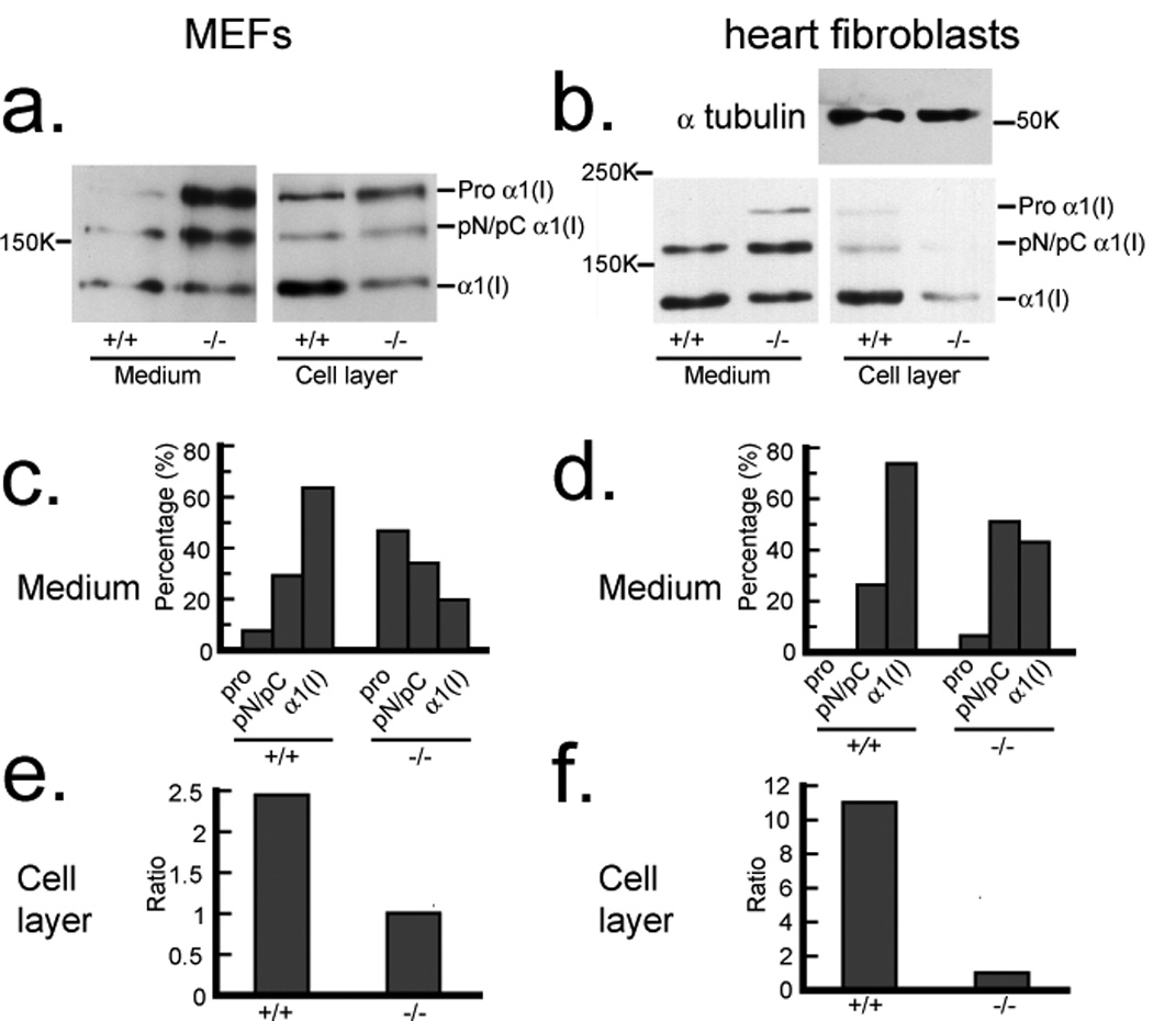
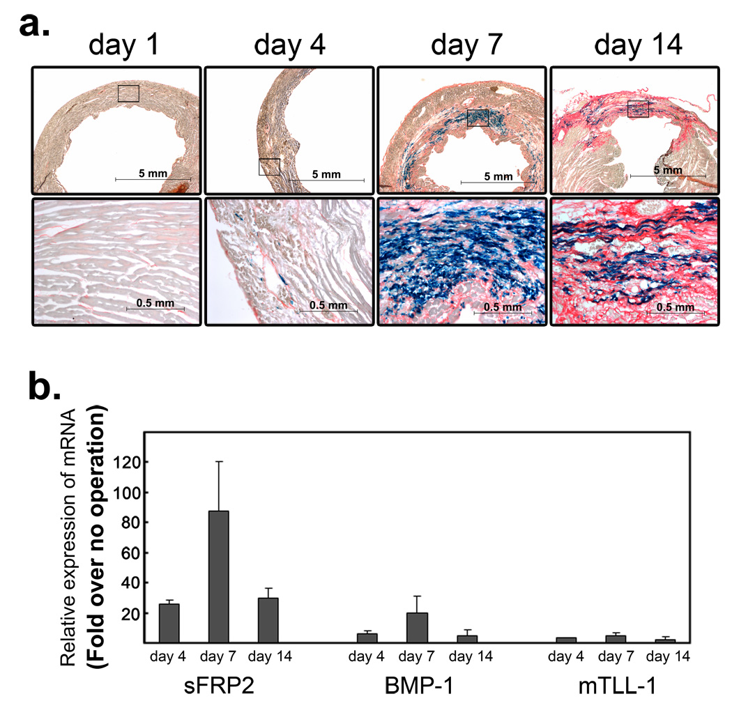
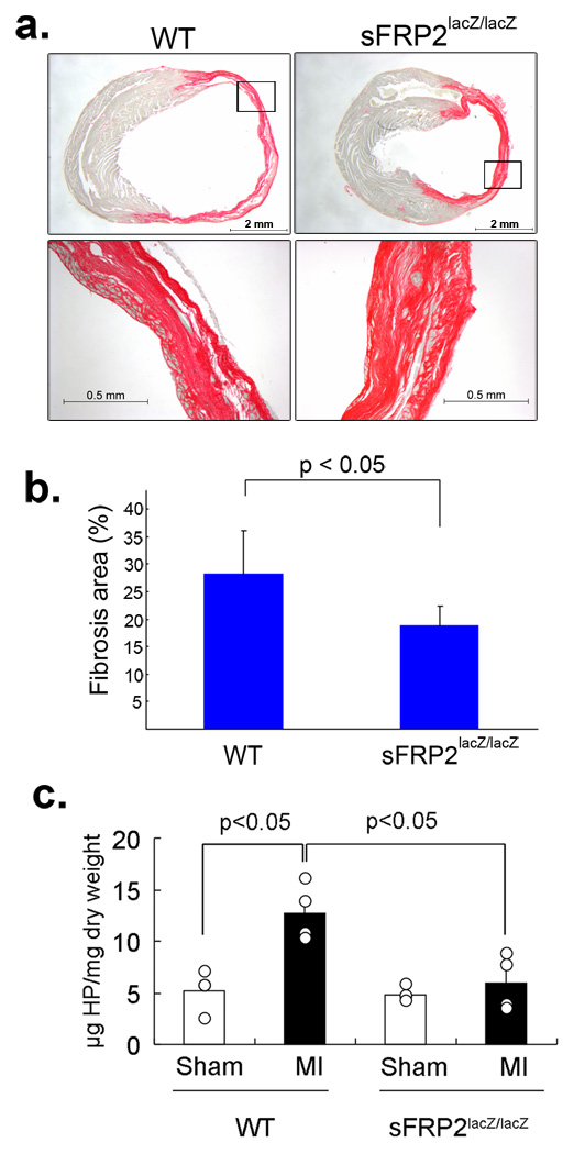
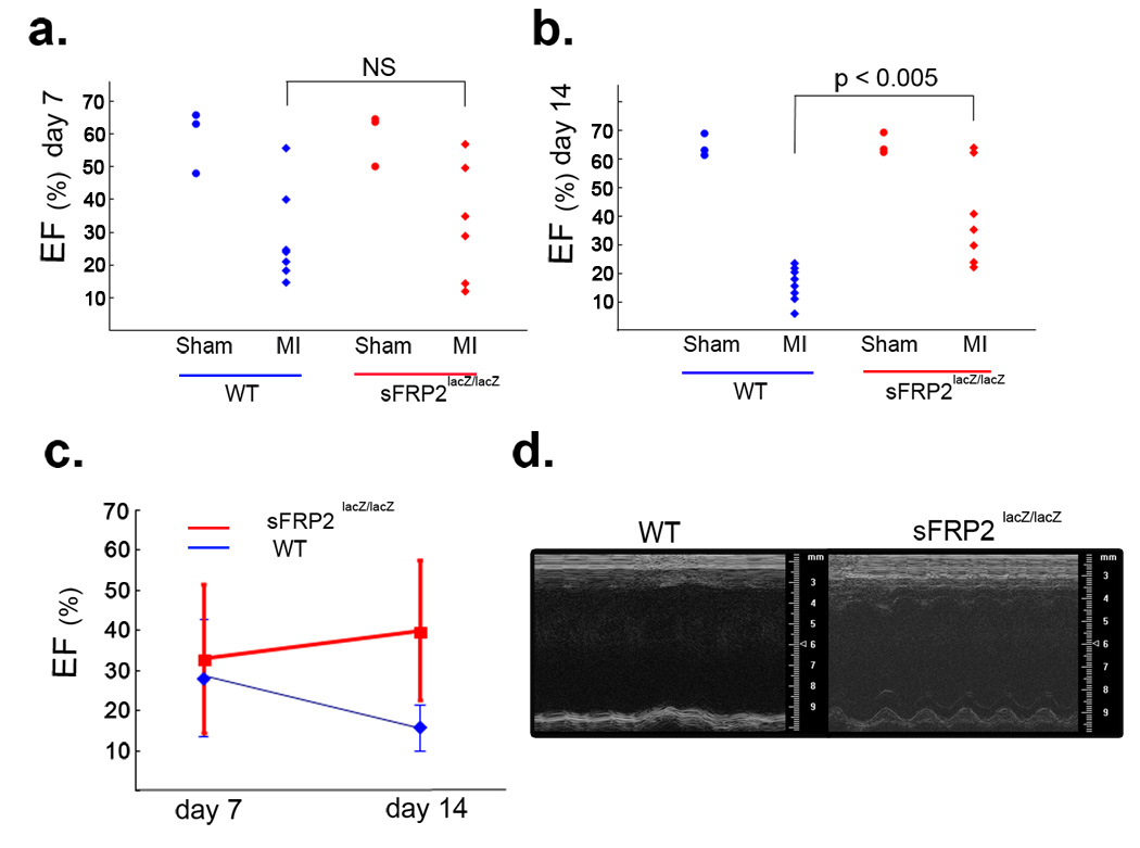
Secreted Frizzled-related proteins (sFRPs) have emerged as key regulators of a wide range of developmental and disease processes. Most of the known functions of mammalian sFRPs have been attributed to their ability to antagonize Wnt signalling. Recently however, Xenopus laevis and zebrafish sFRP, Sizzled, was shown to function as an antagonist of Chordin processing by Tolloid-like metalloproteinases. This has led to the proposal that sFRPs may function as evolutionarily conserved antagonists of chordinase activities of this class of proteinases. In contrast to this proposal, we show here that the mammalian sFRP, sFRP2, does not affect Chordin processing, but instead, can serve as a direct enhancer of procollagen C proteinase activity of Tolloid-like metalloproteinases. We also show that the level of fibrosis, in which procollagen processing by Tolloid-like proteinases has a rate-limiting role, is markedly reduced in Sfrp2-null mice subjected to myocardial infarction. Importantly, this reduced level of fibrosis is accompanied by significantly improved cardiac function. This study thus uncovers a function for sFRP2 and a potential therapeutic application for sFRP2 antagonism in controlling fibrosis in the infarcted heart.


Figures

Figure 1
Figure 1
sFRP2 enhances cleavage of procollagen, but not Chordin, by BMP1 and mTLL1. Immunoblots show absence of effects of increasing concentrations of sFRP2 on the cleavage of Chordin by BMP1 (a) or mTLL1 (b), whereas Szl is shown to efficiently inhibit Chordin cleavage by BMP1 (c), with a much reduced inhibitory effect on Chordin cleavage shown by Szl with an ogon-like D92N mutation (d). Chordin cleavage is evidenced by the appearance of a 15-kDa N-terminal fragment (arrow). In contrast to the absence of sFRP2 effects on Chordin cleavage, autofluorography shows that a 2-fold molar excess of sFRP2 enhanced BMP1 pCP activity, such that no unprocessed pro-forms of the two chain classes of type I procollagen (proα1 and proα2) remained, and greater amounts of pN forms (processing intermediates retaining N-propeptides, but from which C-propeptides have been cleaved) for both chain classes were produced, compared to processing with BMP1 alone (e). Szl inhibits, and SzlD92N has no apparent effect, on BMP1 pCP activity (f). Fold-enhancement of pro-α1(I) to pNα1(I) chain processing by sFRP2 ranged from 1.2 – 2.0 fold in six independent experiments (it is 1.4-fold in fig. 1e) and in the four of these experiments in which pro-α2(I) to pNα2(I) chain ratios could be compared, fold-enhancement of pro-α2(I) processing ranged from 1.2 – 2.6 fold (such a comparison could not be made in fig. 1e, in which no uncleaved proα2(V) chains remained after cleavage of procollagen by BMP1 in the presence or absence of sFRP2). For full scans of a–f, see Supplementary Information, Fig. S6.
Figure 2
Figure 2
sFRP2 binds BMP1 with a KD in the physiological range, predominantly via its Fzl domain. (a) Immunoblots show that His-tagged sFRP2 pulls down FLAG-tagged BMP1, upon precipitation with Ni-NTA agarose. (b) BMP1 binds His-tagged sFRP2 on a BIAcore sensor chip with KD = 7.13 nM. (c) Immunoblots show that the Fc-tagged sFRP2 Fzl domain pulls down FLAG-tagged BMP1 about as effectively as full-length sFRP2-Fc. (d) Fc-tagged Fzl domain of sFRP2 on a Biacore sensor chip binds BMP1 with a KD (KD = 8.68 nM) similar to that of full-length sFRP2-Fc (KD = 6.01 nM), whereas binding of Fc-tagged sFRP2 NTR domain to BMP1 is negligible. (e) Schematic diagram of sFRP2-Fc constructs used for pull-down and BIAcore assays. For full scans of a and c, see Supplementary Information, Fig. S6.
Figure 3
Figure 3
sFRP2 and Szl bind Tolloid-like proteinases via non-protease domain sequences and BMP1/procollagen binding is enhanced by sFRP2. (a) Left, an immunoblot shows input amounts of sFRP2-Fc, FLAG-tagged BMP1 and a deleted form of BMP1 containing all domains except the protease domain (BMP1-D) and isolated BMP1 protease domain (BMP1-P). Right, an immunoblot shows that protein G agarose pull down of sFRP2-Fc pulls down full-length BMP1 and BMP1-D, but not BMP1-P. (b) Left, an immunoblot shows input amounts of sRFP2-Fc and FLAG-tagged mTLL1, or isolated mTLL1 protease domain (mTLL1-P). Right, an immunoblot shows that sFRP2-Fc pulls down full-length mTLL1, but not mTLL1-P. (a and b) arrowheads indicate sFRP2-Fc, and asterisks denote sFRP2 degradation products which bind non-specifically to anti-FLAG antibodies. (c) Left, an immunoblot shows input amounts of His-tagged Szl, and FLAG-tagged BMP1, BMP1-D and BMP1-P. Right, immunoblot shows that Ni-NTA agarose pull down of His-tagged Szl pulls down full-length BMP1 and BMP1-D, but not BMP1-P. (d) Left, an immunoblot shows input amounts of His-tagged Szl, mTLL1, and mTLL1-P. Right, an immunoblot shows that Szl pulls down mTLL1, but not mTLL1-P. (e) Protein domain structures of full-length and truncated forms of BMP1 and mTLL1. Pro, prodomain; Prot, protease domain; CB, CUB domain; E, EGF motif; CT, C-terminal domain. (f) Immunoblots show that procollagen binds sFRP2, and that sFRP2 enhances binding of procollagen to BMP1. Samples were immunoprecipitated with antibody (LF-67) directed against the pro-α1(I) C-telopeptide region. LF-67 is shown to pull down a procollagen/sFRP2-His complex and the presence of sFRP2-His enhances co-immunoprecipitation of procollagen and BMP1*, a form of BMP1 designed to bind but not cleave or release procollagen (Quantification of signals on the blots indicate approximately 4-fold enhancement of BMP1* binding by procollagen in the presence of sFRP2). For full scans of a–d and f, see Supplementary Information, Fig. S6.
Figure 4
Figure 4
Reduced processing of type I procollagen and deposition of collagen into ECM by Sfrp2−/− fibroblasts. Western blot analysis with antibody against the proα1(I) C-telopeptide domain (a small globular region retained on mature α chains, between the triple-helical domain and BMP1 cleavage site) demonstrates decreased processing of procollagen in conditioned media and decreased deposition of collagen into the ECM associated with cell layers of Sfrp2−/− MEF cultures (a) and heart fibroblasts (b) compared to wild-type (sFRP2+/+) cells. Due to the dramatic difference in collagen levels in the cell layer samples of wild-type and Sfrp2−/− heart fibroblasts, samples were re-probed with anti-α tubulin antibody to demonstrate equal loading in the two lanes. Densitometric quantification is shown of the relative amounts of the various collagen forms in conditioned media of MEFs (c) and heart fibroblasts (d). Pro forms are precursors that retain both N- and C-propeptides; α1(I) forms are mature chains, capable of forming fibrils, from which both N- and C-propeptides have been proteolytically removed; pN forms are processing intermediates of proα1(I) chains from which the C- but not N-propeptide has been removed by BMP1-like proteinases; pC forms are processing intermediates from which the N- but not C-propeptide has been removed by non-BMP1-like proteinases. pC and pN forms were not separated by SDS-PAGE in these experiments. Densitometric quantification is shown of the relative amounts of mature α1(I) chains incorporated into cell layer-associated ECM of wild type (+/+) and sFRP2-null (−/−) MEFs (e) and heart fibroblasts (f). For full scans of a and b, see Supplementary Information, Fig. S6.
Figure 5
Figure 5
Induction of Sfrp2 and Bmp1 during the fibrosis phase in the infarcted heart. (a) β-galactosidase staining of the infarcted sFRP2lacZ/+ left-ventricle at days 1, 4, 7, and 14 following coronary artery ligation. Higher magnifications of the boxed areas in the top panels are in bottom panels. At day one, the sirius-red counterstaining (orange) identifies normal collagens in the interstitial space. At this stage, no sFRP2 expression was detected. At day four, the slightly wider areas are stained by sirius-read (orange), which is accompanied by scattered distribution of β-galactosidase-stained sFRP2-expressing fibroblastic cells (blue). By day seven, extensive collagenous fibrosis stained by sirius-red (orange) is detected. Within the fibrotic area, many fibroblastic cells express sFRP2 (blue). At day 14, more extensive sirius-red staining, denoting collagenous scar tissue that has replaced dead myocytes, is clearly detected, and many fibroblastic cells are seen to express sFRP2 (blue). Upregulation of sFRP2 expression in the infarcted area was also confirmed by in situ hybridization staining with a riboprobe for sFRP2 (Supplementary Information: Fig. S3). Scale bars are shown in each panel. (b) Quantitative RT-PCR analysis of sFRP2, BMP1 and mTLL1. Whole ventricle tissue RNA was isolated at days four, seven and 14 following coronary artery ligation. The amounts of sFRP2, BMP1 and mTLL1 were compared to those of non-operated normal ventricle tissue and fold-increases are shown. Duplicate PCR reactions for each sample were carried out and the standard deviations were calculated from the results obtained from two independent experiments. Marked increases of sFRP2 and BMP1 transcripts were detected by day four and their transcript levels continued to increase until day seven. By day 14, their transcript levels decreased, but still remained relatively elevated. The mTLL1 transcript level increase was much less that those of sFRP2 and BMP1. The absolute amounts of sFRP2 transcripts at day 0, day4, day7, day14 were quantified and determined as 6.8×10², 1.5×10⁴, 6.5×10⁴, 1.3×10⁴ molecules per 20ng total RNA, respectively. Expression analysis of all Wnt ligands and Wnt antagonists genes indicates that the expression of Sfrp2 gene exhibits the highest upregulation within the fibrotic area of infarcted hearts (Supplementary Information: Figs. S3&S4).
Figure 6
Figure 6
Reduced fibrosis in the infarcted sFRP2-deficient heart. (a) Sirius-red stained cross sectioned infarcted left ventricle of wild-type (WT) and sFRP2-deficient (Sfrp2lacZ/lacZ) hearts at day 14 after coronary artery ligation. The infarcted wild-type ventricle shows dilatation and extensive sirius-red stained fibrosis (orange). The infarcted Sfrp2lacZ/lacZ ventricle shows much less dilation, and the sirius-red stained area (orange) is reduced. Scale bars are shown in each panel. (b) Quantification of the sirius-red stained fibrotic area. Sirius-red stained areas, determined using AxioVision image analysis software (Carl Zeiss), were compared to whole ventricular areas and are shown as percentages. A representative section from each of a total of nine (n=9) wild type and five (n=5) sFRP2 null hearts was evaluated and the standard deviations were calculated. The sirius-red stained areas in wild type and Sfrp2lacZ/lacZ left ventricles were ∼25 – 35% and ∼15 – 20% of the entire ventricle areas, respectively. (c) Quantification of total hydroxyproline (HP) amounts. Samples from four independent experiments (n=4) for each group were assayed and shown here. The standard deviations and P values are shown. In sham operated control wild type and sFRP2-null hearts, approximately 2 – 5µg of hydroxyproline was present per mg dry weight left-ventricle. In the infarcted wild type heart, approximately 10 – 16µg of hydroxyproline was detected per mg dry weight left-ventricle. In contrast, only 3 – 8µg of hydroxyproline per dry weight left ventricle was detected in the sFRP2-null heart.
Figure 7
Figure 7
Improved cardiac function of the infarcted sFRP2-deficient heart. (a) Ejection fraction (EF) of wild-type (WT) and sFRP2-deficient (Sfrp2lacZ/lacZ) hearts at day seven following coronary artery ligation. Each dot represents an individual mouse (i.e., WT sham: n=3; WT MI: n=7; Sfrp2lacZ/lacZ sham: n=3; Sfrp2lacZ/lacZ MI: n=6). At day seven, the infarcted (MI) hearts of both wild type (WT) and sFRP2-deficient (Sfrp2lacZ/lacZ ) mice showed wide ranges of EF (10% – 55%) and no significant (NS) difference. The EF of sham operated mice showed a normal range of EF. (b) EF of wild-type (WT) and sFRP2-deficient (sFRP2lacZ/lacZ) hearts at day 14 following coronary artery ligation. Each dot represents an individual mouse (i.e., WT sham: n=3; WT MI: n=9; Sfrp2lacZ/lacZ sham: n=3; Sfrp2lacZ/lacZ MI: n=7). At day 14, The infarcted (MI) hearts of all wild type (WT) mice showed consistently low EF (10 – 20%). In contrast, the infarcted (MI) hearts of sFRP2-deficient (Sfrp2lacZ/lacZ) mice showed higher EFs (25 – 60%) and two of the null mice showed EFs (60 –70%) equivalent to those of sham operated mice. The EF of sham operated mice showed a normal range of EF. (c) Change of EFs over time. The EFs of the infarcted wild-type (WT) hearts continued to decline over time, indicating functional failure. In contrast, the EFs of the infarcted sFRP2-deficient (sFRP2lacZ/lacZ) hearts in general remained approximately the same or in some cases slightly improved over time. The data shown in a&b are used to generate this plot. (d) M-mode images of representative wild-type (WT) and sFRP2-deficient (sFRP2lacZ/lacZ) hearts at day 14 following coronary artery ligation. The wild-type heart showed virtually no pumping. In contrast, the sFRP2-deficient heart showed some pumping activity. The time-lapse movie of each heart can be viewed in the (supplementary information Movies 1 & 2).

Comment in

References

    1. Rattner A, et al. A family of secreted proteins contains homology to the cysteine-rich ligand-binding domain of frizzled receptors. Proc Natl Acad Sci U S A. 1997;94:2859–2863. - PMC - PubMed
    1. Mayr T, et al. Fritz: a secreted frizzled-related protein that inhibits Wnt activity. Mech Dev. 1997;63:109–125. - PubMed
    1. Finch PW, et al. Purification and molecular cloning of a secreted, Frizzled-related antagonist of Wnt action. Proc Natl Acad Sci U S A. 1997;94:6770–6775. - PMC - PubMed
    1. Melkonyan HS, et al. SARPs: a family of secreted apoptosis-related proteins. Proc Natl Acad Sci U S A. 1997;94:13636–13641. - PMC - PubMed
    1. Salic AN, Kroll KL, Evans LM, Kirschner MW. Sizzled: a secreted Xwnt8 antagonist expressed in the ventral marginal zone of Xenopus embryos. Development. 1997;124:4739–4748. - PubMed

Publication types

MeSH terms