. 2009 Jan;41(1):118-24.
doi: 10.1038/ng.272. Epub 2008 Dec 14.

Somatic mutations in angiopoietin receptor gene TEK cause solitary and multiple sporadic venous malformations

Affiliations

Somatic mutations in angiopoietin receptor gene TEK cause solitary and multiple sporadic venous malformations

Nisha Limaye et al. Nat Genet. 2009 Jan.

Abstract

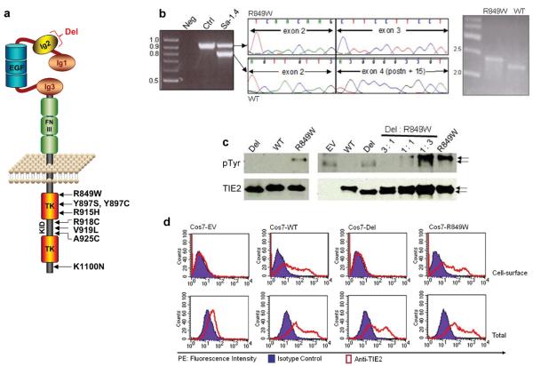
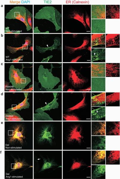
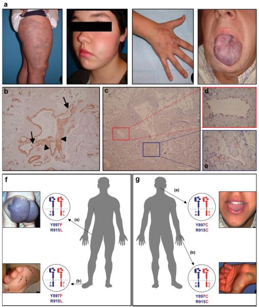
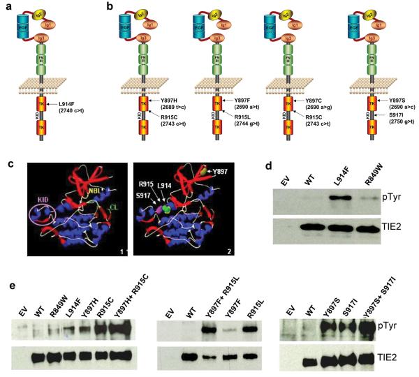
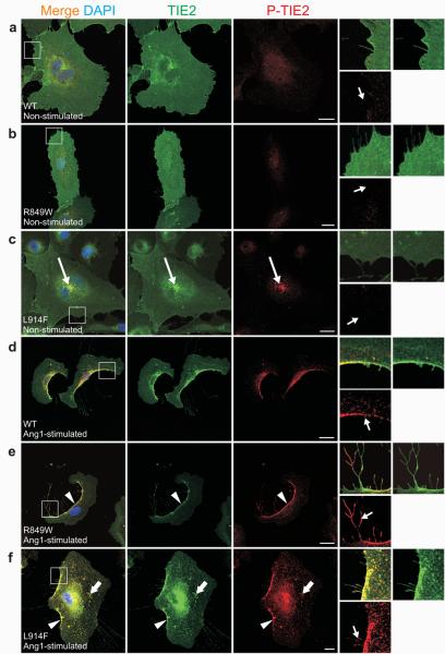
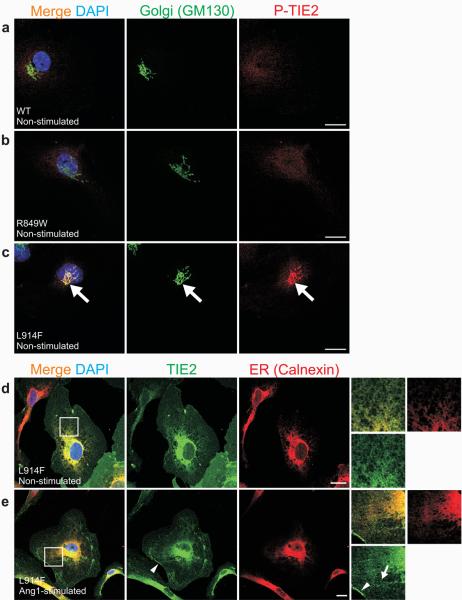
Germline substitutions in the endothelial cell tyrosine kinase receptor TIE2 (encoded by TEK) cause a rare, inherited form of venous anomaly known as a mucocutaneous venous malformation (VMCM; refs. 1, 2, 3 and V.W., N.L., M.U., A. Irrthum, L.M.B. et al., unpublished data). We identified a somatic 'second hit' causing loss of function of TIE2 in a resected VMCM and assessed whether such localized, tissue-specific events have a role in the etiology of sporadic venous malformations, which are far more common. We identified eight somatic TEK mutations in lesions from 28 of 57 individuals (49.1%) with sporadic venous malformations; the mutations were absent from the individuals' blood and control tissues. The somatic mutations included one causing a frequent L914F substitution and several double mutations in cis, all of which resulted in ligand-independent TIE2 hyperphosphorylation in vitro. When overexpressed in human umbilical vein endothelial cells, the L914F mutant was abnormally localized and responded to ligand, in contrast to wild-type TIE2 and the common, inherited R849W mutant, suggesting that the mutations have distinct effects. The presence of the same mutations in multifocal sporadic venous malformations in two individuals suggests a common origin for the abnormal endothelial cells at the distant sites. These data show that a sporadic disease may be explained by somatic changes in a gene causing rare, inherited forms and pinpoint TIE2 pathways as potential therapeutic targets for venous malformations.


Figures

Figure 1
Figure 1. Somatic deletion in Ig2 extracellular domain in a VMCM-tissue
(a) Schematic representation of TIE2: extracellular region with three immunoglobulin-like loops (Ig1, 2, 3), three epidermal growth factor-like repeats (EGF), and three fibronectin type III-like repeats (FNIII). Intracellular region contains 2 tyrosine kinase domains (TK) separated by kinase insert domain (KID). Locations of missense substitutions previously identified in inherited VMCM are shown. A somatic mutation (“Del”) deletes part of Ig2 in the (wild-type) allele of one VMCM patient (Sa-I.4), carrying the inherited R849W mutation. (b) PCR [exons 1-7] in patient Sa-I.4 shows a 900-bp band (expected: 1029 bp) absent in control (left panel). Sequencing demonstrates a 129-bp deletion, of exon 3 and 14-bp of exon 4 (middle) in 900-bp band. Allele-specific PCR shows deletion on wild-type allele (WT), which runs smaller than expected (by 129 bp); mutant R849W runs at expected size (right panel). (c) Western blot with anti-phosphotyrosine antibody to evaluate TIE2 phosphorylation (pTyr, 140 kD, top). Blots stripped and reprobed with anti-TIE2 for total TIE2 levels (bottom). Lysates from Cos-7 cells expressing WT, R849W, or Del (left panel), or various ratios of R849W: Del (right panel). EV: Empty vector control. 43-amino-acid size difference observed in Del, as compared to WT and R849W (arrows). (d) Flow-cytometry on TIE2 (WT, Del, or R849W) -transfected Cos7 cells, or EV controls. Cells stained for cell-surface (upper panels), or total (intracellular + cell surface; lower panels) TIE2. x-axis: Phycoerythrin (PE) fluorescence intensity with anti-TIE2 antibody and mouse IgG1 isotype control; y-axis: cell-counts.
Figure 2
Figure 2. Del-TIE2 is retained in the endoplasmic reticulum (ER)
(a-f) Sparse HUVECs expressing: (a-b) WT, (c-d) R849W, or (e-f) Del mutant-forms of TIE2, stimulated for one hour with Ang1 (b, d and f), or left unstimulated (a, c and e), and stained with antibodies against TIE2 and ER-specific chaperone Calnexin. Magnifications of boxed areas are shown on right. In unstimulated WT and R849W cells, TIE2 is evenly distributed on plasma membranes. Light accumulation of TIE2, with variation between cells, observed in perinuclear area, likely representing newly synthesized or recycled receptors (a and c, middle panels). Ang1-stimulation induced clustering of TIE2-receptors (b and d, arrowheads in insets) and translocation to the retracting cell edges (b and d, arrowheads in middle panels). Del-TIE2 is accumulated in ER in both unstimulated and Ang1-stimulated cells (e-f). Consistent with ER-location, soluble Ang1 failed to induce Del-TIE2 clustering as seen in WT and R849W; only dot-like clusters observed in cell margins, likely representing endogenous TIE2 (arrow). Scale bar 20 μm.
Figure 3
Figure 3. Typical features of sporadic venous malformations
(a) Lesion on leg, cheek, hand, and tongue of patients #11, 19, 13, and 4, respectively. All have the L914F mutation. (b-e) Immunohistochemical staining of sections of dysmorphic vessels and surrounding areas, from VM of patient # 15, with L914F. (b) Smooth muscle alpha-actin demonstrates variable smooth muscle cell layers, thin (arrow) and thick (arrowhead). (c) TIE2 stains a continuous, thin layer of endothelial cells (ECs), shown at higher magnification (boxed) in (d) and (e). TIE2 expression is not detected outside endothelial cells. (f, g) Double-mutant TIE2 alleles in patients with multifocal VMs. (f) Patient #27: large buttock-VM at birth, appearance of small (<1 cm diameter) lesions (round, hyperkeratotic when on plantar surface) by age 6; [Y897F + R915L] TIE2 cis double-mutations in both buttock and foot lesions. (g) Patient #28: multiple (sub)cutaneous and intramuscular lesions in the face, thorax and abdominal regions, with new lesions appearing by age 18. Tongue and wrist VMs, both with [Y897C + R915C]. Normal allele, blue; mutant, red.
Figure 4
Figure 4. Somatic TIE2 mutations identified in sporadic VM tissues cause ligand-independent hyperphosphorylation
(a-b) Schematic representations of TIE2 showing the missense substitutions, and corresponding amino acid changes identified in sporadic VMs. Amino acid residues 897, 914 and 915 located in first TK, and 917 in KID. (a) L914F: most frequent change, 24/28 patients. (b) In 4 patients, TIE2 double-mutations occur in cis. [Y897F+R915L] and [Y897C+R915C] in 2 lesions each, from 2 patients with multifocal VMs. (c) Representation of TIE2 intracellular region protein structure (Panel 1, KID = Kinase Insert Domain, NBL = Nucleotide Binding Loop, CL = Catalytic Loop), showing the positions of the somatic mutations (Panel 2). (d,e) Western blot with anti-phosphotyrosine antibody to evaluate TIE2 phosphorylation (pTyr, 140 kD, top). Blots stripped and reprobed with anti-TIE2 antibody to detect total TIE2 levels (bottom). (d) L914F compared to wild-type TIE2 (WT) and common germline mutant R849W. (e) Comparison of double mutants (Y897H+R915C, Y897F+R915L, Y897S+S917I) with WT and constituent single-mutants. EV: empty vector controls.
Figure 5
Figure 5. Ang1-induced TIE2 clustering and phosphorylation in HUVECs expressing mutant (R849W and L914F) and wild-type (WT) TIE2
(a-f) Sparse HUVECs expressing: (a, d) WT, (b, e) R849W, or (c, f) L914F, stimulated for one hour with Ang1 (d-f), or left unstimulated (a-c), and stained with antibodies against total TIE2 (TIE2) and phosphorylated TIE2 (P-TIE2). Boxed areas magnified on right. (a-c) In unstimulated WT and R849W expressing cells, TIE2 distributed evenly on cell membranes with no P-TIE2 specific staining. In contrast, increased L914F immunostaining observed in perinuclear area, where L914F is activated based on P-TIE2 staining (c, long arrows). Without Ang1, neither cell edges nor cellular extensions showed P-TIE2 antibody reactivity (small arrows). (d-f) In response to Ang1-stimulation, TIE2 translocated and phosphorylated in cell rear (arrowheads) and in retraction fibers (small arrows, right). Ang1 promotes WT translocation and activation in cell rear (d). R849W showed enhanced phosphorylation in retraction fibers (e, small arrow) when compared to WT (d, small arrow). Ang1 induced punctuate L914F clustering (thick arrows), and less complete translocation to cell rear (f). Scale bar 20 μm.
Figure 6
Figure 6. Cellular localization and activation of L914F-TIE2
(a-e) Sparse HUVECs expressing: (a) WT, (b) R849W or (c-e) L914F forms of TIE2 stimulated for one hour with Ang1 (e), or left unstimulated (a-d), and stained with antibodies against Golgi-specific GM130 and phospho-TIE2 (P-TIE2; a-c), or total TIE2 (TIE2) and ER-specific Calnexin (d-e). (a-c) When compared to WT (a) and R849W (b), L914F showed ligand-independent activation in the Golgi apparatus (c, large arrows). (d-e) In both unstimulated and Ang1-stimulated cells, L914F was found to localize in ER and plasma-membrane. Ang1 induced L914F clustering and translocation, albeit incompletely, to retracting cell edges (e, arrowhead). TIE2 clusters in L914F-expressing cells typically appeared punctuate (e, arrow). Scale bar 20 μm.

References

    1. Vikkula M, et al. Vascular dysmorphogenesis caused by an activating mutation in the receptor tyrosine kinase TIE2. Cell. 1996;87:1181–90. - PubMed
    1. Calvert JT, et al. Allelic and locus heterogeneity in inherited venous malformations. Hum Mol Genet. 1999;8:1279–89. - PubMed
    1. Wouters V, Boon LM, Mulliken JB, Miikka Vikkula. TIE2 and Cutaneomucosal Venous Malformation. In: Epstein C, Erickson RP, Wynshaw-Boris A, editors. Inborn Errors of development. 2nd. Oxford Univeristy Press Inc.; 2007.
    1. Wouters V, Limaye N, Uelbelhoer M, Irrthum A, Boon LM, Mulliken JB, Enjolras O, Baselga E, Berg J, Dompmartin A, Ivarsson SA, Kangesu L, Lacassie Y, Murphy J, Teebi AA, Pennington A, Rieu P, Vikkula M. Hereditary Cutaneomucosal Venous Malformations Are Caused by Variably Activating TIE2 Mutations. submitted.
    1. Dumont DJ, Yamaguchi TP, Conlon RA, Rossant J, Breitman ML. tek, a novel tyrosine kinase gene located on mouse chromosome 4, is expressed in endothelial cells and their presumptive precursors. Oncogene. 1992;7:1471–80. - PubMed

Publication types

MeSH terms

Associated data