. 2009 Jan;16(1):17-22.
doi: 10.1038/nsmb.1534. Epub 2008 Dec 14.

Histone H3 tail clipping regulates gene expression

Affiliations

Histone H3 tail clipping regulates gene expression

Helena Santos-Rosa et al. Nat Struct Mol Biol. 2009 Jan.

Abstract

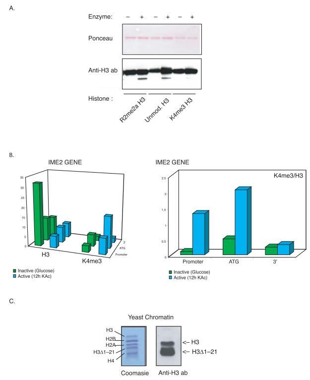
Induction of gene expression in yeast and human cells involves changes in the histone modifications associated with promoters. Here we identify a histone H3 endopeptidase activity in Saccharomyces cerevisiae that may regulate these events. The endopeptidase cleaves H3 after Ala21, generating a histone that lacks the first 21 residues and shows a preference for H3 tails carrying repressive modifications. In vivo, the H3 N terminus is clipped, specifically within the promoters of genes following the induction of transcription. H3 clipping precedes the process of histone eviction seen when genes become fully active. A truncated H3 product is not generated in yeast carrying a mutation of the endopeptidase recognition site (H3 Q19A L20A) and gene induction is defective in these cells. These findings identify clipping of H3 tails as a previously uncharacterized modification of promoter-bound nucleosomes, which may result in the localized clearing of repressive signals during the induction of gene expression.


Figures

Figure 1
Figure 1
A histone H3 endopeptidase activity in S. cerevisiae. (A) A H3 endopeptidase activity is present in the yeast nuclei. Nuclear extracts from early exponential, sporulation or stationary phase cultures were assayed for endopeptidase activity on recombinant H3. The reactions were stained with Ponceau (15 μl reaction, to visualize the products) and analyzed by western blot with anti C-terminal H3 antibody (5 μl reaction, to avoid signal saturation). The clipped H3 product is highlighted. (B) The H3 endopeptidase activity is enriched upon nutrient starvation. Extracts from early exponential, sporulation or stationary phase cultures, purified on sepharose beads, were assayed for endopeptidase activity on calf H3. The reactions were analyzed by western blot with anti C-terminal H3 antibody. The clipped H3 product is highlighted. (C) The Q19L20A21 is the recognition sequence for the H3 endoppetidase. Extract from yeast cell on stationary phase was pull down on Sepharose beads and assayed against recombinant wild type H3 and recombinant Q19L20->AA mutant H3.The reactions were analysed by western blot with anti C-terminal H3 antibody. (D) Activity against different histones. Stationary phase pull down on Sepharose beads was assayed against identical amounts of calf histones. The reactions were analysed by western blot with antibodies specific for each histone. The clipped H3 product is highlighted.
Figure 2
Figure 2
The histone H3 endopeptidase is a serine protease with preference for non-activating marks. (A) Histone H3 tails carrying non activatory marks are preferentially clipped. Histone H3 modified at different residues (see Materials and Methods) was used as substrate for the endopeptidase activity pulled down from stationary phase culture. The reactions were analyzed by western blot with anti C-terminal H3 antibody. (B) K4 me3 H3 are not evicted from promoters upon gene induction. Chromatin immunoprecipitation experiments were performed in yeast cells cultured in either glucose (green bars) or sporulation medium for 12 hours (blue bars) using anti K4 me3 H3 antibody and anti C-terminal H3 antibody. The precipitated DNA was analyzed by quantitative PCR using primers specific to the indicated positions within the IME2 gene. The diagrams represent relative fluorescent units normalized to an intergenic region on chromosome V (see Material and Methods). (C) H3Δ1-21 occurs in vivo in S. cerevisiae. Yeast chromatin was prepared from early stationary phase culture and analyzed by Coomasie (left panel) or western blot (right panel). The full length and clipped H3 (H3Δ1-21) are highlighted.
Figure 3
Figure 3
The H3 endopeptidase is a serine protease. (A) Inhibition of the H3 endopeptidase activity. The endopeptidase activity on recombinant H3 was tested in the presence of different proteases inhibitors. The reactions were analysed by Western Blot with anti C-terminal H3 antibody. (B) Protease inhibitor profile of the H3 endopeptidase activity.
Figure 4
Figure 4
Histone tails are removed prior core depletion from promoters. (A) Histone H3 tails are removed specifically from promoters. Chromatin immunoprecipitation experiments were performed in yeast cells cultured in either glucose (green bars) or sporulation medium (read and blue bars) using anti-myc antibody to detect the N-terminus of histone H3 (N) and anti C-terminal H3 antibody to detect the H3 core (C). The precipitated DNA was analyzed by quantitative PCR using primers specific to the indicated positions within the gene. The diagrams represent relative fluorescent units normalized to an intergenic region on chromosome V (see Material and Methods). (B) Histone H3 tails are removed on induction of the sporulation. Left panel: Chromatin immunoprecipitation experiments were performed in yeast cells cultured in either glucose (green bars) or sporulation medium (read and blue bars) using anti-myc antibody to detect the N-terminus of histone H3 (N) and anti C-terminal H3 to detect the H3 core (C). The precipitated DNA was analyzed by quantitative PCR using primers specific to the indicated promoters. The diagrams represent relative fluorescent units normalized to an intergenic region on chromosome V. Right panel: RT-PCR analysis was performed on the same cultures described (left panel), using primers specific to the indicated genes. The expression level of each gene was normalized to the RNA levels of RTG2. (C) Histone H3 tails are removed on the transition to stationary phase. Left panel: Chromatin immunoprecipitation experiments were performed in yeast cells cultured in glucose to OD600nm: 0.6 (green bars), OD600nm: 5 (read bars), OD600nm: 11 (blue bars) using anti-myc antibody to detect the N-terminus of histone H3 (N) and anti C-terminal H3 to detect the H3 core (C). The precipitated DNA was analyzed by quantitative PCR using primers specific to the indicated genes. The diagrams represent relative fluorescent units normalized to an intergenic region on chromosome V. Right panel: RT-PCR analysis was performed on the same cultures described (left panel), using primers specific to the indicated genes. The expression level of each gene was normalized to the RNA levels of RTG2.
Figure 5
Figure 5
Histone H3 Q19L20->AA mutation abrogates tail loss and cause transcription defects. (A) The H3 Q19L20->AA yeast strain does not loose H3 tails. Nuclear fractions from wild type and Q19L20->AA H3 yeast strains cultured in glucose to OD600nm: 7 were analyzed by western blot with anti C-terminal H3 antibody. The clipped H3 product is highlighted. (B) Gene induction is defective in the H3 Q19L20->AA yeast strain. RT-PCR analysis was performed on cultures of wild type and Q19L20->AA H3 grown on rich medium to OD600nm: 0.6 and OD600nm: 7, using primers specific to the indicated genes. The histogram bar represent folds induction for each strain and gene, calculated as the ration of [induced mRNA: non induced mRNA].

Similar articles

Cited by

References

    1. Kouzarides T. Chromatin modifications and their function. Cell. 2007;128:693–705. - PubMed
    1. Lee CK, Shibata Y, Rao B, Strahl BD, Lieb JD. Evidence for nucleosome depletion at active regulatory regions genome-wide. Nat Genet. 2004;8:900–905. - PubMed
    1. Korber P, Luckenbach T, Blaschke D, H^rz W. Evidence for histone eviction in trans upon induction of the yeast PHO5 promoter. Mol Cell Biol. 2004;24:10965–10974. - PMC - PubMed
    1. Bernstein BE, Liu CL, Humphrey EL, Perlstein EO, Schreiber SL. Global nucleosome occupancy in yeast. Genome Biol. 2004;5:R62. - PMC - PubMed
    1. Schermer UJ, Korber P, H^rz W. Histones are incorporated in trans during reassembly of the yeast PHO5 promoter. Mol Cell. 2005;19:279–85. - PubMed

MeSH terms