. 2009 Jan;30(1):90-7.
doi: 10.1038/aps.2008.7. Epub 2008 Dec 15.

Transformation and action of extracellular NAD+ in perfused rat and mouse livers

Affiliations

Transformation and action of extracellular NAD+ in perfused rat and mouse livers

Ana Carla Broetto-Biazon et al. Acta Pharmacol Sin. 2009 Jan.

Abstract

Aim: Transformation and possible metabolic effects of extracellular NAD+ were investigated in the livers of mice (Mus musculus; Swiss strain) and rats (Rattus novergicus; Holtzman and Wistar strains).

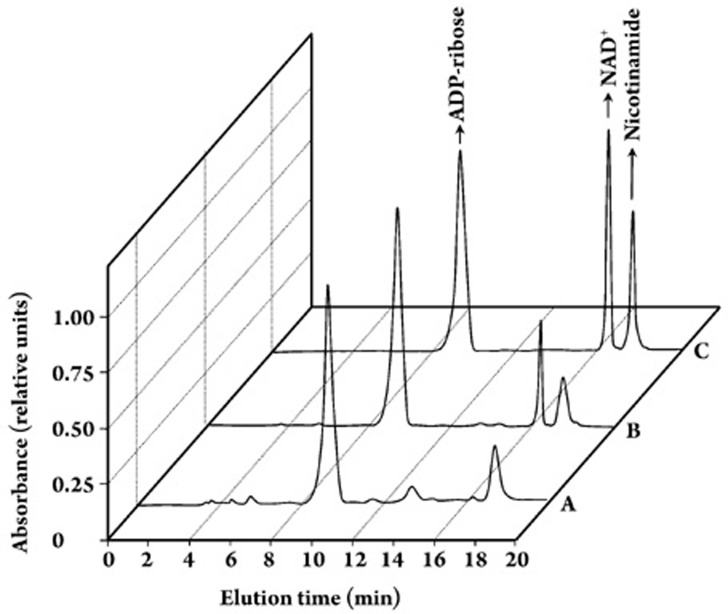
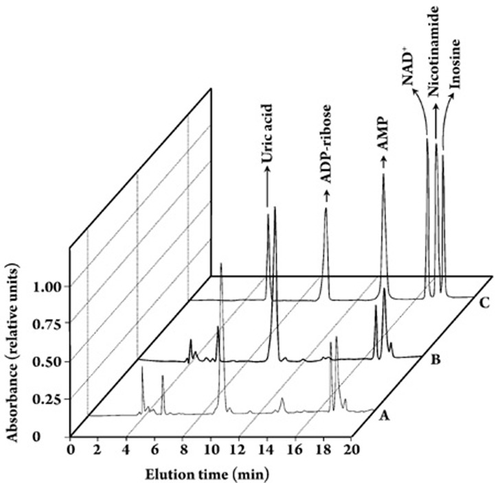
Methods: The livers were perfused in an open system using oxygen-saturated Krebs/Henseleit-bicarbonate buffer (pH 7.4) as the perfusion fluid. The transformation of NAD+ was monitored using high-performance liquid chromatography.

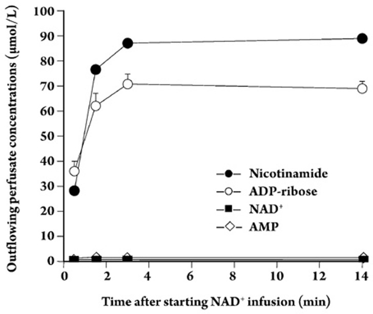
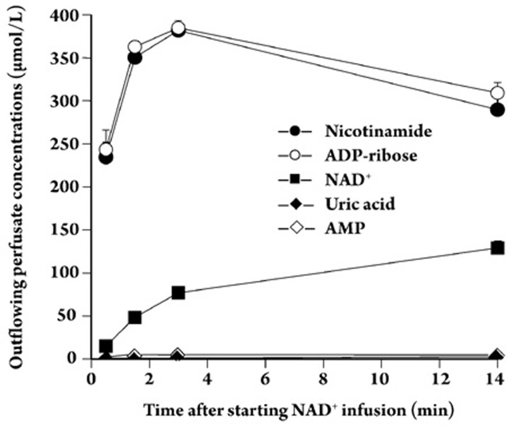
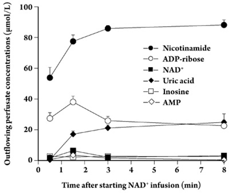
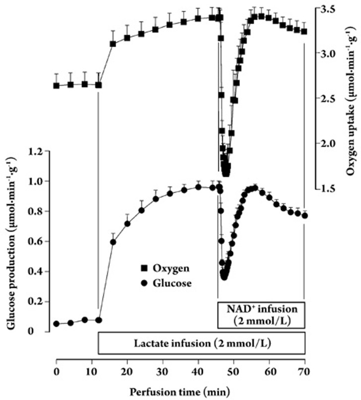
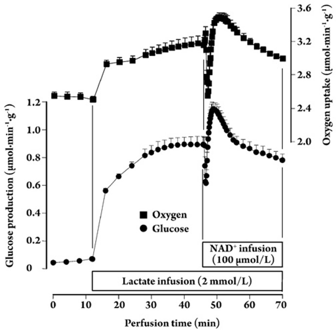
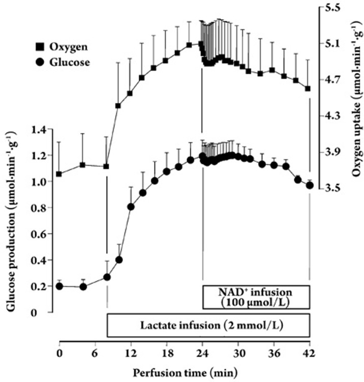
Results: In the mouse liver, the single-pass metabolism of 100 micromol/L NAD+ was almost complete; ADP-ribose and nicotinamide were the main products in the outflowing perfusate. In the livers of both Holtzman and Wistar rats, the main transformation products were ADP-ribose, uric acid and nicotinamide; significant amounts of inosine and AMP were also identified. On a weight basis, the transformation of NAD+ was more efficient in the mouse liver. In the rat liver, 100 micromol/L NAD+ transiently inhibited gluconeogenesis and oxygen uptake. Inhibition was followed by a transient stimulation. Inhibition was more pronounced in the Wistar strain and stimulation was more pronounced in the Holtzman strain. In the mouse liver, no clear effects on gluconeogenesis and oxygen uptake were found even at 500 micromol/L NAD+.

Conclusion: It can be concluded that the functions of extracellular NAD+ are species-dependent and that observations in one species are strictly valid for that species. Interspecies extrapolations should thus be made very carefully. Actually, even variants of the same species can demonstrate considerably different responses.


Figures

Figure 1
Figure 1
Typical chromatographic profiles of standards and outflowing per-fusate of the mouse liver. Perfusate samples were collected at 1.3 min after initiation of NAD+ infusion. HPLC fractionation with spectrophotometric detection at 254 nm was done as described in the Materials and methods section. Legends: (A) outflowing perfusate with 100 μmol/L NAD+ infusion; (B) outflowing perfusate with 500 μmol/L NAD+ infusion; (C) standards. The absorbance scale was normalized as a fraction of the highest absorbance peak in each run.
Figure 2
Figure 2
Typical chromatographic profiles of standards and outflowing perfusate of rat livers from the Wistar and Holzman strains. Perfusate samples were collected at 1.3 min after initiation of 100 μmol/L NAD+ infusion. HPLC fractionation with spectrophotometric detection at 254 nm was done as described in the Materials and methods section. Legends: (A) Holzman strain; (B) Wistar strain; (C) standards. The absorbance scale was normalized as a fraction of the highest absorbance peak in each run.
Figure 3
Figure 3
Time course of 100 μmol/L NAD+ transformation in the perfused mouse liver. Livers of fasted mice were perfused with Krebs/Henseileit-bicarbonate buffer (pH 7.4) as described in Materials and Methods. The concentrations of NAD+ and its metabolites in the outflowing perfusate at different times after initiation of NAD+ infusion were determined by HPLC. Data are the means±mean standard errors of four liver perfusion experiments.
Figure 4
Figure 4
Time course of 500 μmol/L NAD+ transformation in the perfused mouse liver. Livers of fasted mice were perfused with Krebs/Henseleit-bicarbonate buffer (pH 7.4) as described in Materials and Methods. The concentrations of NAD+ and its metabolites in the outflowing perfusate at different times after initiation of NAD+ infusion were determined by HPLC. Data are the means±mean standard errors of four liver perfusion experiments.
Figure 5
Figure 5
Time course of 100 μmol/L NAD+ transformation in the perfused liver of Holtzman rats. Livers of fasted rats were perfused with Krebs/Henseleit-bicarbonate buffer (pH 7.4) as described in Materials and Methods. The concentrations of NAD+ and its metabolites in the outflowing perfusate at different times after initiation of NAD+ infusion were determined by HPLC. Data are the means±mean standard errors of four liver perfusion experiments.
Figure 6
Figure 6
Gluconeogenesis and oxygen uptake in the liver of Wistar rats: influence of lactate and NAD+. Livers from 24-h fasted rats were perfused as described in Materials and Methods. After oxygen uptake stabilization, lactate and NAD+ (at the given concentrations) were infused at the indicated times. Samples of the effluent perfusate were collected for glucose assay. Oxygen was monitored polarographically. Data represent the means±SEM of five liver perfusion experiments.
Figure 7
Figure 7
Gluconeogenesis and oxygen uptake in the liver of Holtzman rats: influence of lactate and NAD+. Livers from 24-h fasted rats were perfused as described in Materials and Methods. After oxygen uptake stabilization, lactate and NAD+ (at the given concentrations) were infused at the indicated times. Samples of the effluent perfusate were collected for glucose assay. Oxygen was monitored polarographically. Data represent the means±SEM of five liver perfusion experiments.
Figure 8
Figure 8
Gluconeogenesis and oxygen uptake in the mouse liver: influence of lactate and NAD+. Livers from 24-h fasted mice were perfused as described in Materials and Methods. After oxygen uptake stabilization, lactate and NAD+ (at the given concentrations) were infused at the indicated times. Samples of the effluent perfusate were collected for glucose assay. Oxygen was monitored polarographically. Data represent the means±mean standard errors of five liver perfusion experiments.

Similar articles

Cited by

References

    1. Liersch M, Grotelüschen H, Decker K. NAD permeation into the liver cell. Hoppe-Seyler's Z Physiol Chem. 1971;352:267–74. - PubMed
    1. Ziegler M. New functions of a long-known molecule. Emerging roles of NAD in cellular signaling. Eur J Biochem. 2000;267:1550–64. - PubMed
    1. Rusinko N, Lee HC. Widespread occurrence in animal tissues of an enzyme catalyzing the conversion of NAD+ into a cyclic metabolite with intracellular Ca2+-mobilizing activity. J Biol Chem. 1989;264:11725–31. - PubMed
    1. Chini EN, Klener P, Jr, Beers KW, Chini CCS, Grande JP, Dousa TP. Cyclic ADP-ribose metabolism in rat kidney: high capacity for synthesis in glomeruli. Kidney Int. 1997;51:1500–6. - PubMed
    1. Khoo KM, Chang CF. Localization of plasma membrane CD38 is domain specific in rat hepatocyte. Arch Biochem Biophys. 2000;373:35–43. - PubMed

Publication types