. 2009 Jan;30(1):98-106.
doi: 10.1038/aps.2008.11. Epub 2008 Dec 15.

Inhibition of tubulointerstitial fibrosis by pentoxifylline is associated with improvement of vascular endothelial growth factor expression

Affiliations

Inhibition of tubulointerstitial fibrosis by pentoxifylline is associated with improvement of vascular endothelial growth factor expression

Qiu-gen Zhou et al. Acta Pharmacol Sin. 2009 Jan.

Abstract

Aim: Recent information indicates that pentoxifylline (PTX) has the ability to suppress inflammation and profibrotic cell proliferation. In this study, we investigated the effect of PTX on tubulointerstitial fibrosis and the expression of vascular endothelial growth factor (VEGF) in a rat model of obstructive nephropathy.

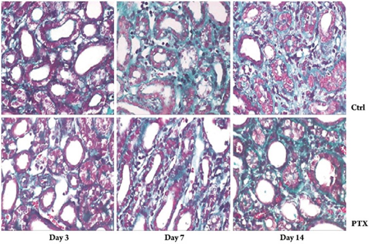
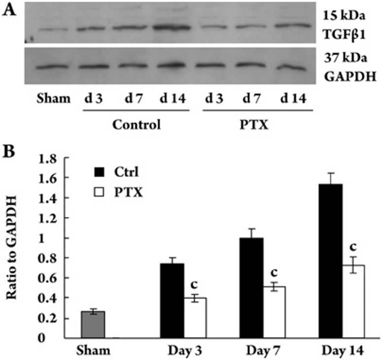
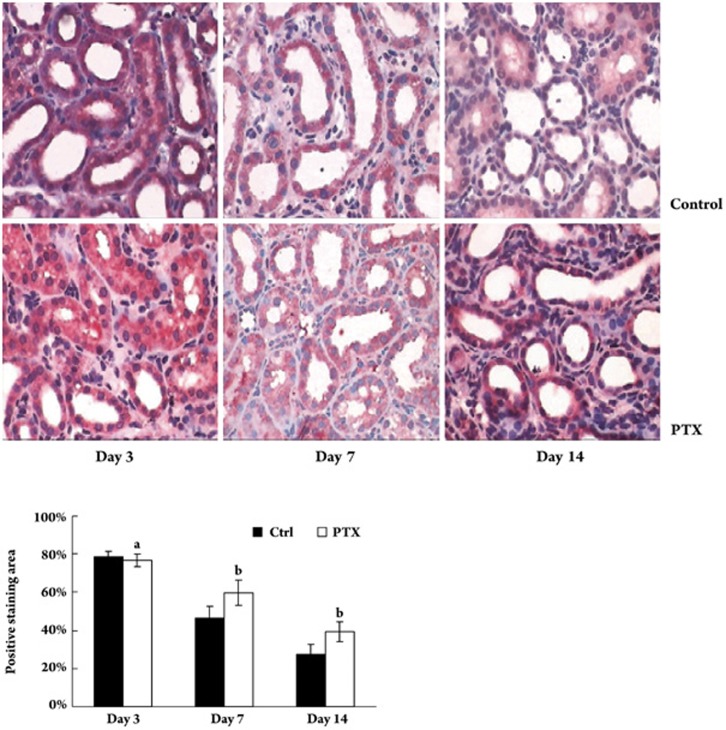
Methods: Wistar rats with left ureteral ligation were divided into control and PTX-treated groups. The histopathologic degree of tubulointerstitial fibrosis was scored with PAS and Masson-stained sections. The protein and mRNA for vascular endothelial growth factor (VEGF) were semiquantitatively measured with immunohistochemistry and RT-PCR. The protein for transforming growth factor beta1 (TGFbeta1) and hypoxia-induced factor 1 alpha (HIF-1alpha) was determined by Western blot.

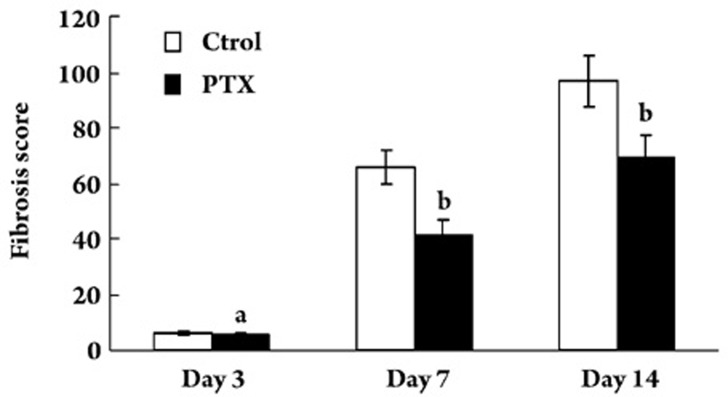
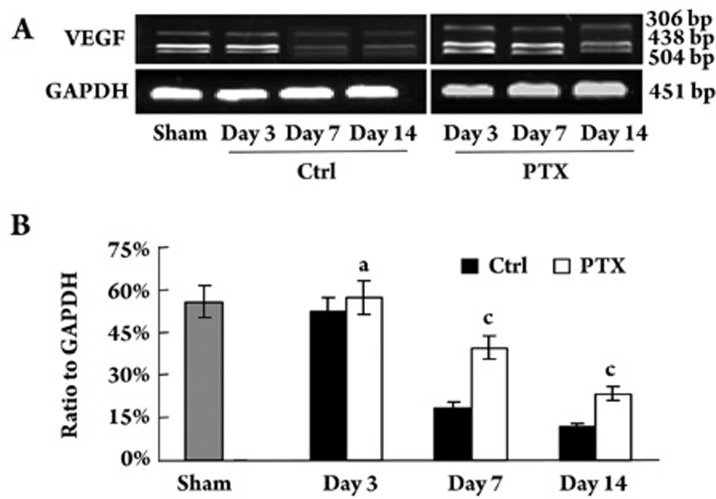
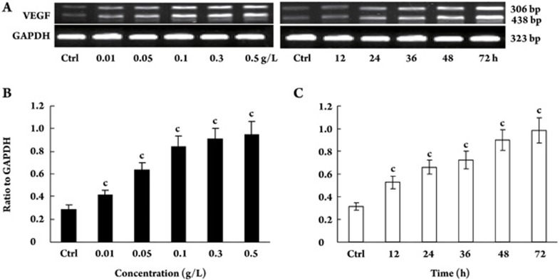
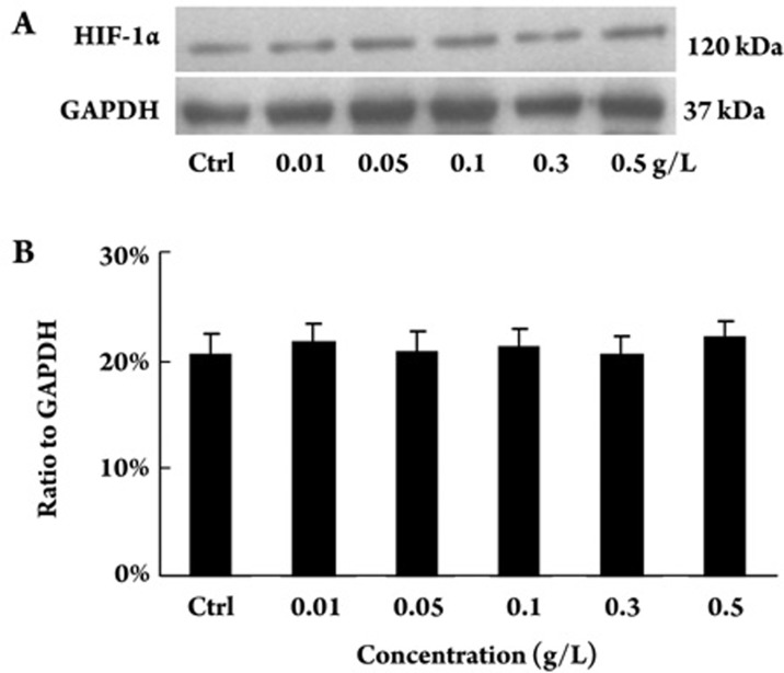
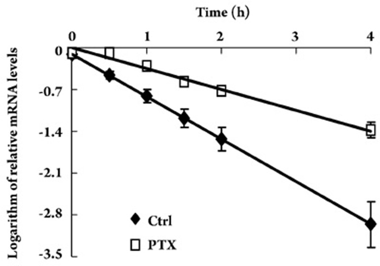
Results: Compared with the control group, PTX treatment reduced fibrosis scores at d 7 and d 14 (P<0.05). The reduction was accompanied by inhibited expression of transforming growth factor-beta 1 (TGFbeta1), a key cytokine in tubulointerstitial fibrogenesis (P<0.01). Meanwhile, VEGF protein and mRNA in the kidney were increased in the PTX-treated group compared with the control group (P<0.01). PTX up-regulated expression of VEGF mRNA in a dose- and time-dependent manner in cultured HK-2 cells (P<0.01). However, expression of HIF-1alpha (a key transcription factor for VEGF gene expression) was unchanged by PTX treatment. PTX prolonged the half-life of VEGF mRNA by a 1.07-fold increase.

Conclusions: PTX inhibited tubulointerstitial fibrosis in a rat model of obstructive nephropathy while preventing loss of VEGF. PTX up-regulated expression of VEGF mRNA through stabilization of its mRNA in cultured renal tubular epithelial cells.


Figures

Figure 1
Figure 1
Representative histopathology in UUO rats treated with or without PTX (Massone stain, ×400). Day 3 after ligation, renal tubular dilation with flattened epithelium and without atrophy or thickness of tubular basement membrane accompanied with few fibrosis in interstitial space. Day 7 after ligation, some tubular atrophy and thickness of tubular basement membrane accompanied with moderate fibrosis in interstitial space. Day 14 after ligation, diffused tubular atrophy and thickness of tubular basement membrane accompanied with severe fibrosis in interstitial space.
Figure 2
Figure 2
Semiquantitative tubulointerstitial fibrosis score in control and PTX-treated group. Compared with control group, the fibrosis score was reduced in PTX treated group at day 7 and day 14. The values were expressed as mean±SD of 3 independent experiments. aP>0.05, bP<0.05 vs control group.
Figure 3
Figure 3
Expression of TGFβ1 protein in kidney tissue. (A) Representative western blot of TGFβ1 in sham and UUO rat treated with or without PTX. (B) Compared with control group, PTX treatment inhibited the increased expression of TGFβ1. The data was shown as ratio of TGFβ1 density to GAPDH density. The values were expressed as mean±SD of 3 independent experiments. cP<0.01 vs control.
Figure 4
Figure 4
Expression of VEGF protein in the kidney determined with immunohistochemistry in UUO rats treated with or without PTX. Compared with control group, PTX treatment improved the reduced expression of VEGF at day 7 and day 14. Data were shown as percentage of the staining area to selected field and expressed as mean±SD of 3 independent experiments. aP>0.05, bP<0.05 vs control group.
Figure 5
Figure 5
Expression of VEGF mRNA in the kidney determined with RT-PCR in UUO rats treated with or without PTX. (A) Representative electrophoresis of RT-PCR products. VEGF120, VEGF164, and VEGF188 of 3 VEGF isoforms were expressed in the kidney. (B) Compared with control group, PTX treatment improved the reduced expression of VEGF mRNA at day 7 and day 14. The band intensities corresponding to the 306 bp, 438 bp, and 504 bp mRNAs were quantified. Data were shown as ratio of VEGF density to that of GAPDH and expressed as mean±SD of 3 independent experiments. aP>0.05, cP<0.01 vs control group.
Figure 6
Figure 6
Effect of PTX on expression of VEGF mRNA in cultured HK-2 tubular epithelial cells. (A) Representative electrophoresis of RT-PCR products. VEGF121 and VEGF165 of 2 VEGF isoforms were expressed in cultured HK-2 tubular epithelial cells. (B) Dose-dependent effect of PTX on VEGF mRNA expression. HK-2 cells were treated with indicated concentration of PTX for 72 h. The VEGF mRNA was semiquantatively determined with RT-PCR. The band intensities corresponding to the 306 bp and 438 bp mRNAs were quantified. The data were shown as ratio of VEGF density to that of GAPDH and expressed as mean±SD of 3 independent experiments. (C) Time-dependent effect of PTX on VEGF mRNA expression. HK-2 cells were treated with 0.5 g/L PTX for the indicated time. cP<0.01 vs control by ANOVA.
Figure 7
Figure 7
Effect of PTX on expression of HIF-1α in cultured HK-2 tubular epithelial cells. HK-2 cells were treated with indicated concentration of PTX for 72 h. The expression of HIF-1α was determined with Western blot. (A) Representative Western blot of HIF-1α in HK-2 cells. (B) Compared with control, PTX treatment did not change the expression of HIF-1α. The data were shown as ratio of HIF-1α density to that of GAPDH and expressed as mean±SD of 3 independent experiments. P=0.935 vs control by ANOVA.
Figure 8
Figure 8
Effect of PTX on VEGF mRNA stability. Overnight serum deprived HK-2 cells were treated with PTX of 0.5 g/L for 24 h before treatment with 5 μmol/L actinomycin D. VEGF mRNA levels at 0, 0.5, 1, 1.5, 2, and 4 h after addition of actinomycin D were assessed by RT-PCR. The half-life (T1/2) of VEGF mRNA was calculated from the slope of the plot of versus time of decay, using linear regression analysis. The estimated half-life of VEGF mRNA was 0.715±0.094 h. PTX treatment prolonged the half-life with increase by 1.07 fold. P<0.01 vs control.

Similar articles

Cited by

References

    1. Damico G, Ferrario F, Rastaldi MP. Tubulointerstitial damage in glomerular disease: its role in the progression of renal damage. Am J Kidney Dis. 1995;26:124–32. - PubMed
    1. Kang DH, Kanellis J, Hugo C, Truong L, Anderson S, Kerjaschki D, et al. Role of the microvascular endothelium in progressive renal disease. J Am Soc Nephrol. 2002;13:806–16. - PubMed
    1. Nangaku M. Chronic hypoxia and tubulointerstitial injury: A final common pathway to end-stage renal failure. J Am Soc Nephrol. 2006;17:17–25. - PubMed
    1. Choi YJ, Chakraborty S, Nguyen V, Nguyen C, Kim BK, Shim SI, et al. Peritubular capillary loss is associated with chronic tubulointerstitial injury in human kidney: Altered expression of vascular endothelial growth factor. Hum Pathol. 2000;31:1491–7. - PubMed
    1. Ohashi R, Kitamura H, Yamanaka N. Peritubular capillary injury during the progression of experimental glomerulonephritis in rats. J Am Soc Nephrol. 2000;11:47–56. - PubMed

Publication types

MeSH terms

Substances