. 2009 Feb;89(2):131-41.
doi: 10.1038/labinvest.2008.120. Epub 2008 Dec 15.

Keratinocyte-derived follistatin regulates epidermal homeostasis and wound repair

Affiliations

Keratinocyte-derived follistatin regulates epidermal homeostasis and wound repair

Maria Antsiferova et al. Lab Invest. 2009 Feb.

Abstract

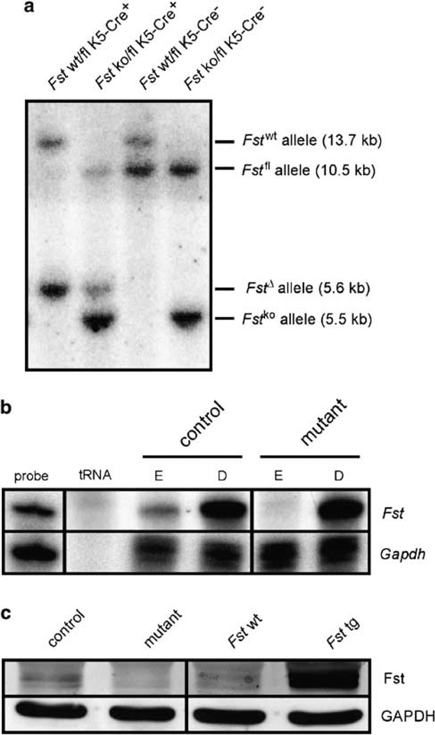
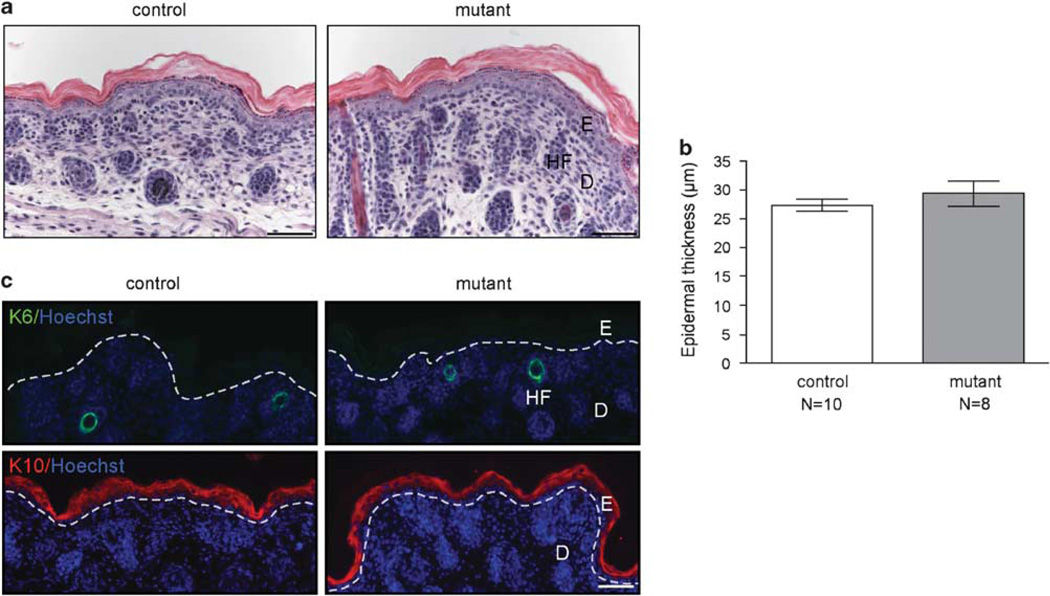
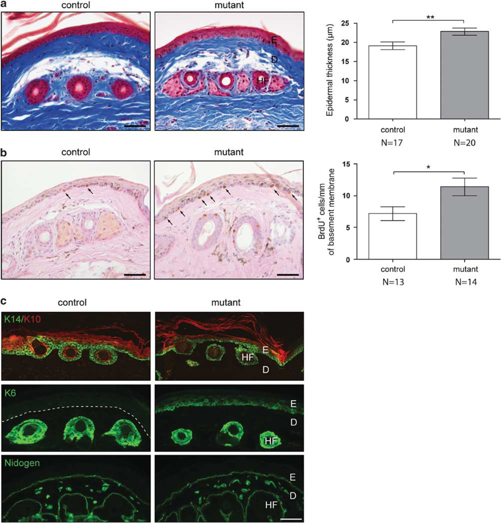
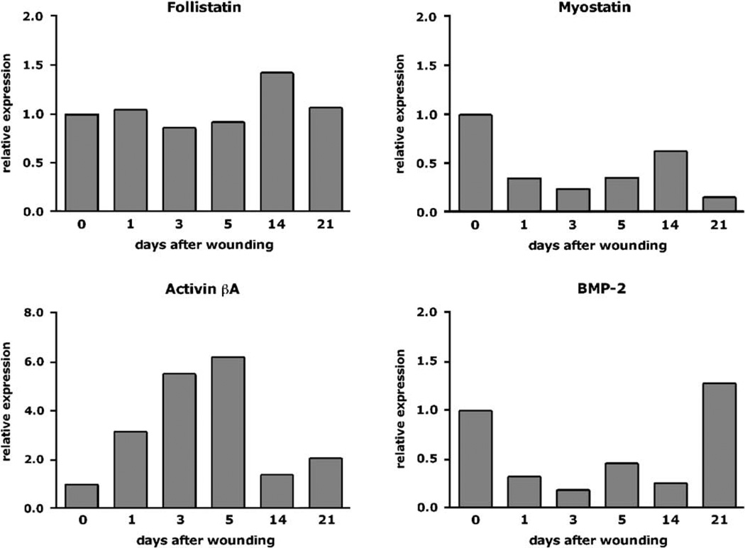
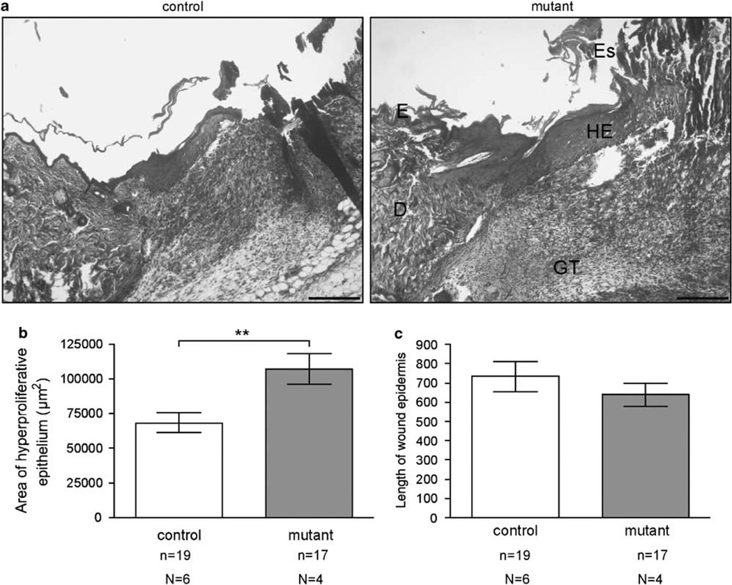
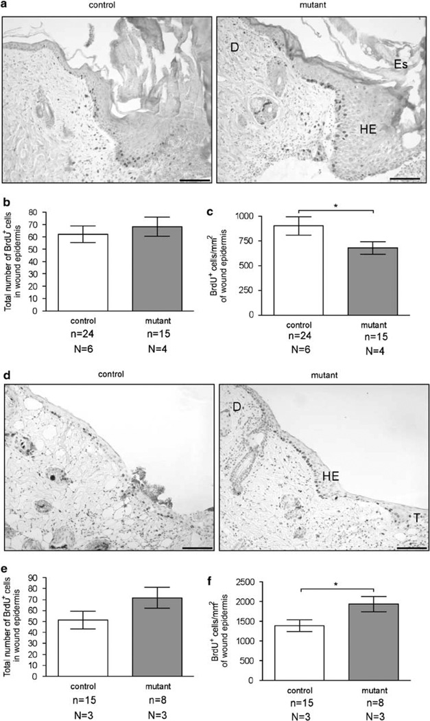
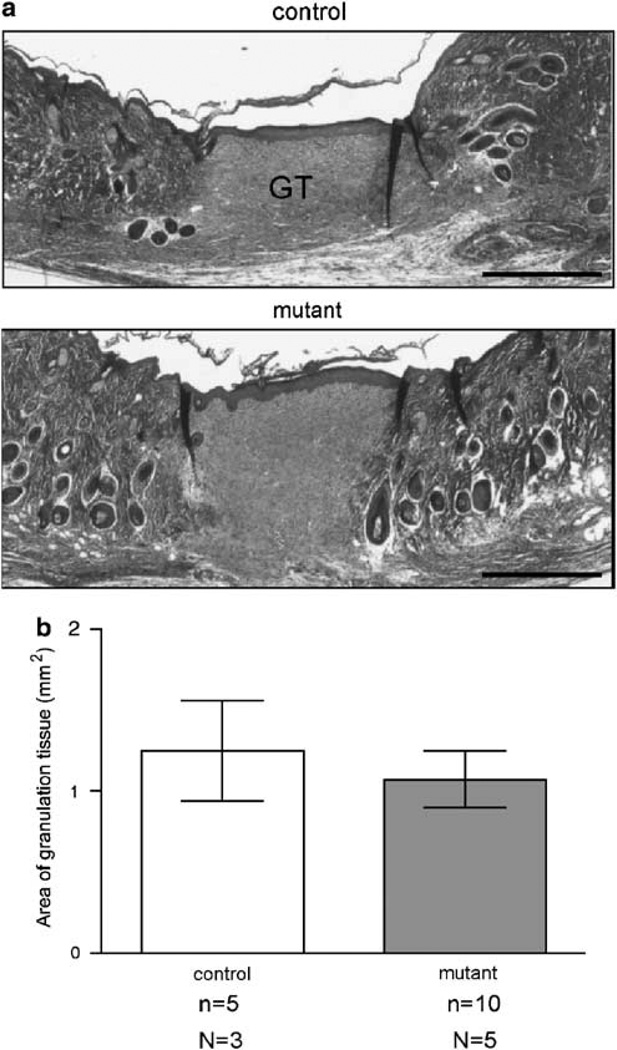
Activin is a growth and differentiation factor that controls development and repair of several tissues and organs. Transgenic mice overexpressing activin in the skin were characterized by strongly enhanced wound healing, but also by excessive scarring. In this study, we explored the consequences of targeted activation of activin in the epidermis and hair follicles by generation of mice lacking the activin antagonist follistatin in keratinocytes. We observed enhanced keratinocyte proliferation in the tail epidermis of these animals. After skin injury, an earlier onset of keratinocyte hyperproliferation at the wound edge was observed in the mutant mice, resulting in an enlarged hyperproliferative epithelium. However, granulation tissue formation and scarring were not affected. These results demonstrate that selective activation of activin in the epidermis enhances reepithelialization without affecting the quality of the healed wound.


Figures

Figure 1
Figure 1
Efficient knockout (ko) of follistatin in the epidermis. (a) Southern blot analysis showing Cre-mediated deletion of the floxed (fl) Fst allele. A 1230 bp SacI/HindIII probe was used to detect wild-type (wt), ko, fl and deleted (Δ) Fst alleles in genomic DNA digested with SacI and EcoRI. (b) Lack of follistatin mRNA in the epidermis is shown by RNase protection assay. Follistatin mRNA is expressed in the dermis and the epidermis of control mice but only in the dermis of Fst mutant mice. Samples of 20 µg of total RNA were used for hybridization. The hybridization probes (1000 c.p.m.) served as size markers (probe). tRNA (10 µg) was used as a negative control. Hybridization of RNA with a GAPDH riboprobe served as a loading control. (c) Western blot analysis showing lack of follistatin in primary keratinocytes isolated from mutant mice. Cell lysates were prepared from primary keratinocytes isolated from mutant (Fst ko/fl K5Cre+) and control (Fst wt/fl K5Cre) littermates. Protein (100 µg) was analyzed by western blot analysis with an antibody against follistatin. Protein lysates of primary keratinocytes isolated from mice overexpressing follistatin in the epidermis (Fst tg) served as a positive control. Expression of follistatin in the wt littermates of these transgenic mice is also shown for comparison (Fst wt).
Figure 2
Figure 2
Lack of skin abnormalities in newborn Fst mutant mice. (a) Masson trichrome-stained histological sections of newborn control and Fst mutant skin are shown. (b) Morphometric analysis of tail skin revealed no difference in epidermal thickness. N =number of mice. Results shown are mean ± s.e.m. (c) Expression of keratin 6 (K6; first panel, green) and keratin 10 (K10; second panel, red) is similar in control and mutant mice. Dotted line indicates epidermal–dermal border. (a, c) Bar = 50µm. E: epidermis; D: dermis; HF: hair follicle.
Figure 3
Figure 3
Lack of follistatin in keratinocytes affects skin homeostasis of adult mice. (a) Masson trichrome-stained histological sections of tail skin of adult control and Fst mutant mice are shown. Morphometric analysis of tail skin revealed a significant increase in epidermal thickness in Fst mutant mice (**P=0.0093). (b) Increased number of BrdU-positive keratinocytes in tail skin of mutant mice. Arrows indicate stained nuclei. BrdU-positive cells per mm of basement membrane were counted (*P =0.0263). (c) Expression of keratin 14 (K14; first panel, green) and keratin 10 (K10; first panel, red) is similar in control and mutant mice. Fst mutant mice show interfollicular expression of keratin 6 (K6; second panel). Nidogen expression is unaltered in mutant mice (third panel). Dotted line indicates epidermal–dermal border. (a–c) Bar = 50 µm. E: epidermis; D: dermis; HF: hair follicle. (a, b) N = number of mice. Results shown are mean ± s.e.m.
Figure 4
Figure 4
Expression of follistatin and its binding partners in healing skin wounds. RNA was isolated from normal and wounded skin at different stages after injury and analyzed by quantitative RT-PCR for the mRNA levels of follistatin and its binding partners. Expression levels of follistatin, activin βA, myostatin and BMP-2 were normalized using GAPDH and RNA polymerase II polypeptide A as reference genes. Expression levels in unwounded skin (0) were set as 1. Each RNA sample was a pool from at least eight wounds.
Figure 5
Figure 5
Enlarged area of hyperproliferative epithelium 5 days after wounding in Fst mutant mice. (a) Masson trichrome-stained histological sections of 5-day wounds (5 dw) of control and mutant mice are shown. E: epidermis; D: dermis; HE: hyperproliferative epithelium; GT: granulation tissue; Es: eschar. Bar = 200 µm. (b, c) Morphometric analysis of wound healing parameters. The area of the hyperproliferative epithelium in 5 dw is significantly increased in Fst mutant mice (b: **P = 0.0049), whereas the length of the wound epidermis is similar in control and mutant mice (c). n = number of measurements; N = number of mice. Results shown are mean±s.e.m.
Figure 6
Figure 6
Increased proliferation of keratinocytes at the wound edge 3 days after wounding. (a, d) Detection of proliferating cells in the hyperproliferative epithelium 5 (a) and 3 days after wounding (d). HE: hyperproliferative epithelium; D: dermis; Es: eschar; T: tip of migrating epithelial tongue. Bar= 100 µm. (b, c, e, f) Quantification of proliferating cells in 5-day (b, c) and 3-day (e, f) wounds. The total number of BrdU-positive cells in the hyperproliferative epithelium (b, e) and the number of BrdU-positive cells per area of hyperproliferative epithelium (c, f) are shown. n= number of measurements; N = number of mice (c: *P= 0.0499; f: * = 0.0421). Results shown are mean±s.e.m.
Figure 7
Figure 7
Reepithelialization, keratinocyte differentiation and granulation tissue formation are unaltered in Fst mutant mice 13 days after wounding. (a) Masson trichrome-stained histological sections of 13-day wounds (13 dw) of control and mutant mice are shown. GT: granulation tissue. Bar= 500 µm. (b) The area of granulation tissue in 13 dw is similar in control and mutant mice. n = number of measurements; N = number of mice. Results shown are mean±s.e.m.

References

    1. Phillips DJ. The activin/inhibin family. In: Thomson AW, Lotze MT, editors. The Cytokine Handbook. 4th edn. London: Academic Press; 2003. pp. 1153–1177.
    1. Mathews LS. Activin receptors and cellular signaling by the receptor serine kinase family. Endocr Rev. 1994;15:310–325. - PubMed
    1. Nakamura T, Takio K, Eto Y, et al. Activin-binding protein from rat ovary is follistatin. Science. 1990;247:836–838. - PubMed
    1. Chang H, Brown CW, Matzuk MM. Genetic analysis of the mammalian transforming growth factor-beta superfamily. Endocr Rev. 2002;23:787–823. - PubMed
    1. Matzuk MM, Lu N, Vogel H, et al. Multiple defects and perinatal death in mice deficient in follistatin. Nature. 1995;374:360–363. - PubMed

Publication types

MeSH terms