. 2009 Jan;2(1):33-42.
doi: 10.1038/mi.2008.62. Epub 2008 Sep 17.

Guanylate-binding protein-1 is expressed at tight junctions of intestinal epithelial cells in response to interferon-gamma and regulates barrier function through effects on apoptosis

Affiliations

Guanylate-binding protein-1 is expressed at tight junctions of intestinal epithelial cells in response to interferon-gamma and regulates barrier function through effects on apoptosis

M Schnoor et al. Mucosal Immunol. 2009 Jan.

Abstract

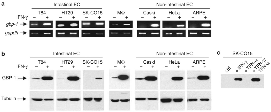
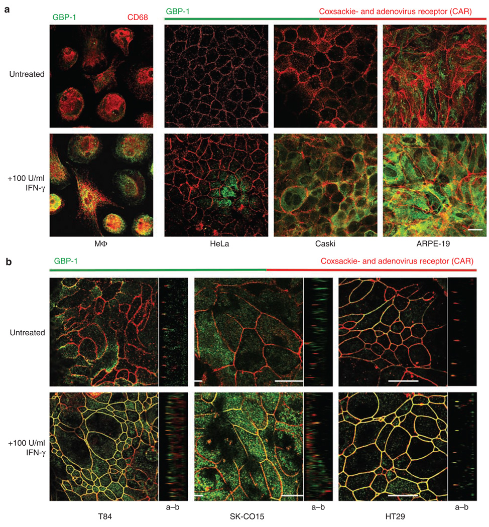
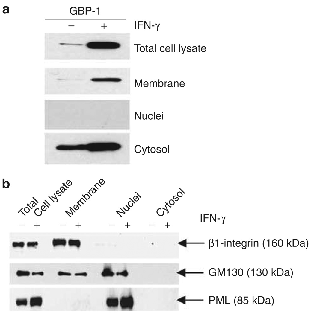
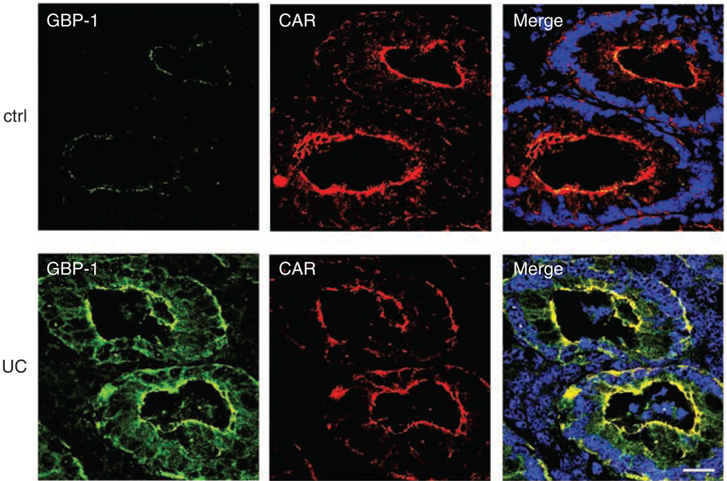
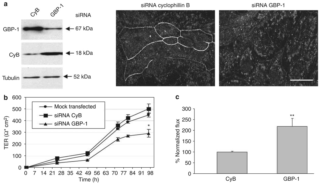
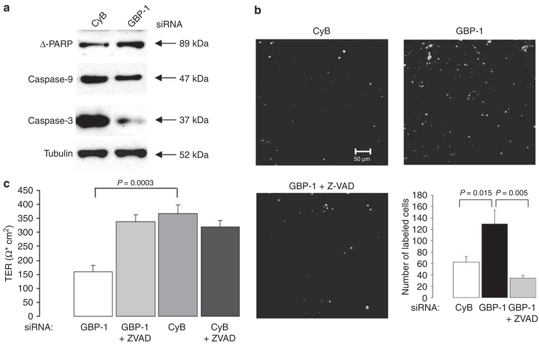
Guanylate-binding protein-1 (GBP-1) is an interferon inducible large GTPase involved in endothelial cell proliferation and invasion. In this report, expression and function of GBP-1 were investigated in vitro in intestinal epithelia after exposure to interferon-gamma and in human colonic mucosa from individuals with inflammatory bowel disease (IBD). Interestingly, in contrast to other epithelia, GBP-1 distributed to the plasma membrane in intestinal epithelial cells where it colocalized with the tight junction protein coxsackie- and adenovirus receptor. In addition, expression of GBP-1 was upregulated in colonic epithelia of individuals with IBD. Downregulation of GBP-1 by siRNA resulted in enhanced permeability that correlated with increased apoptosis. Indeed, inhibition of caspase activity prevented the inhibition of barrier formation induced by the loss of GBP-1. These data suggest that GBP-1 is a novel marker of intestinal mucosal inflammation that may protect against epithelial apoptosis induced by inflammatory cytokines and subsequent loss of barrier function.


Figures

Figure 1
Figure 1
Human epithelial cell lines express guanylate-binding protein-1 (GBP-1) mRNA and protein after interferon (IFN)-γ treatment. (a) Reverse transcriptase (RT)–PCR analysis of GBP-1 expression. Intestinal as well as nonintestinal epithelial cell (EC) lines show strong expression of GBP-1 mRNA after IFN-γ treatment for 24 h. Macrophage (MΦ) cDNA was used as a positive control, glyceraldehyde 3-phosphate dehydrogenase as loading control. (b) Western blot analysis of GBP-1 expression. All EC lines tested show strong production of GBP-1 protein after IFN-γ treatment. MΦ cell lysates served as positive control and tubulin blots as loading control. (c) Tumor necrosis factor (TNF)-α increases the susceptibility of SK-CO15 cells to IFN-γ. However, TNF-α alone does not induce GBP-1 expression. Displayed are representative images of three independent PCR and western blot experiments.
Figure 2
Figure 2
Guanylate-binding protein-1 (GBP-1) is localized at tight junctions (TJ) in intestinal epithelial cells after interferon (IFN)-γ treatment. (a) Immunofluorescence (IF) staining of MΦ and nonintestinal epithelial cell lines show a strong increase of GBP-1 expression after IFN-γ treatment. However, GBP-1 is only detected in the cytosol. (b) In intestinal epithelial cells, GBP-1 is not only induced after IFN-γ treatment but a great portion of the produced GBP-1 can be detected at intercellular junctions colocalizing with the TJ molecule coxsackie- and adenovirus receptor (CAR). Images of the xz axes corroborate TJ localization of GBP-1 in intestinal epithelial cell lines (ab : apical–basal). Bar = 20 µm.
Figure 3
Figure 3
Guanylate-binding protein-1 (GBP-1) cosediments with membrane fractions after treatment with interferon (IFN)-γ. (a) Cellular fractionation of untreated or IFN-γ-treated T84 cells and western blot analysis revealed that GBP-1 is present in the membrane fraction of IFN-γ-treated cells. (b) Western blots confirmed the purity of the fractions. β1-Integrin was only detected in the membrane fraction and promyelocytic leukemia protein (PML) only in the nuclear fraction. The absence of β1-integrin and the Golgi marker 130 (GM130) from the cytosolic fraction confirmed that the cytosolic fraction contained no membrane residues.
Figure 4
Figure 4
Guanylate-binding protein-1 (GBP-1) is upregulated in colonic mucosa of individuals with ulcerative colitis (UC). Human colon tissue sections (n = 3) of individuals with active UC or Crohn’s disease and control tissue (ctrl) without inflammation were labeled with monoclonal GBP-1 (green) and coxsackie- and adenovirus receptor (CAR) (red) antibodies. Very little GBP-1 is expressed in the control tissue, whereas UC tissue shows a strong staining of GBP-1 in crypt epithelium in colocalization with CAR. The displayed image of the UC tissue is representative for the results obtained with all inflammatory bowel disease (IBD) tissue samples. Nuclei are shown in blue. Bar = 20 µm.
Figure 5
Figure 5
Guanylate-binding protein-1 (GBP-1) downregulation inhibits epithelial barrier formation. (a) Downregulation of GBP-1 by siRNA is efficient and specific in SK-CO15 cells treated with interferon (IFN)-γ and tumor necrosis factor (TNF)-α. Western blot analyses show silencing of GBP-1 in cells transfected with GBP-1-specific siRNA, whereas cells transfected with siRNA specific for cyclophilin B (CyB) show no unspecific silencing. Tubulin is shown as a loading control. The immunofluorescent (IF) image shows specific removal of GBP-1 from the lateral cell membrane. Bar = 20µm. (b) SK-CO15 cells were transfected with siRNA specific for CyB or GBP-1 after 24 h exposure to IFN-γ and TNF-α and transepithelial electrical resistance (TER) was measured every 24 h in five independent experiments (n = 5). Cells transfected with GBP-1 siRNA showed significantly lower TER compared with cells transfected with CyB siRNA 96 h after transfection (*P < 0.05). (c) SK-CO15 cells were transfected with siRNA specific for CyB or GBP-1. After differences in TER were measured fluorescein isothiocyanate (FITC)-labeled 4kDa dextran was added to the apical side and 24 h later the flux was fluorimetrically assessed in four independent experiments (n = 4). Paracellular flux was increased more than twofold in cells transfected with GBP-1 siRNA compared with cells transfected with CyB siRNA (**P < 0.01).
Figure 6
Figure 6
Guanylate-binding protein-1 (GBP-1) downregulation promotes apoptosis. (a) Apoptosis was analyzed by western blot using different markers (poly-(ADP-ribose)-polymerase (PARP), caspases 3 and 9). GBP-1 silencing increases the susceptibility of SK-CO15 cells to apoptosis as all, PARP and procaspases 3 and 9 are cleaved to a greater extent compared with cells transfected with cyclophilin B (CyB) siRNA. Tubulin was used as a loading control. (b) TUNEL staining of SK-CO15 cells transfected with either CyB- or GBP-1-specific siRNA. The images shown are representative fields of the stained filters. The graph represents quantification of data derived from five different fields. SK-CO15 cells transfected with GBP-1 siRNA show a more than twofold increase in TUNEL staining compared with controls. When treated with the caspase inhibitor Z-VAD, these cells are protected from apoptosis. (c) Cells transfected with either CyB or GBP-1 siRNA were treated with Z-VAD or left untreated. Z-VAD treatment prevents the inhibition of TER development in cells transfected with GBP-1 siRNA, whereas control cells are not affected by Z-VAD treatment.

Similar articles

Cited by

References

    1. Cheng YS, Colonno RJ, Yin FH. Interferon induction of fibroblast proteins with guanylate-binding activity. J. Biol. Chem. 1983;258:7746–7750. - PubMed
    1. Naschberger E, Bauer M, Sturzl M. Human guanylate-binding protein-1 (hGBP-1) characterizes and establishes a nonangiogenic endothelial cell activation phenotype in inflammatory diseases. Adv. Enzyme Regul. 2005;45:215–227. - PubMed
    1. Olszewski MA, Gray J, Vestal DJ. In silico genomic analysis of the human and murine guanylate-binding protein (GBP) gene clusters. J. Interferon Cytokine Res. 2006;26:328–352. - PubMed
    1. Prakash B, Praefcke GJ, Renault L, Wittinghofer A, Herrmann C. Structure of human guanylate-binding protein-1 representing a unique class of GTP-binding proteins. Nature. 2000;403:567–571. - PubMed
    1. Guenzi E, et al. The guanylate-binding protein-1 GTPase controls the invasive and angiogenic capability of endothelial cells through inhibition of MMP-1 expression. EMBO J. 2003;22:3772–3782. - PMC - PubMed

Publication types

MeSH terms