Non-natural and photo-reactive amino acids as biochemical probes of immune function
- PMID: 19079589
- PMCID: PMC2592539
- DOI: 10.1371/journal.pone.0003938
Non-natural and photo-reactive amino acids as biochemical probes of immune function
Abstract
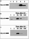
Wilms tumor protein (WT1) is a transcription factor selectively overexpressed in leukemias and cancers; clinical trials are underway that use altered WT1 peptide sequences as vaccines. Here we report a strategy to study peptide-MHC interactions by incorporating non-natural and photo-reactive amino acids into the sequence of WT1 peptides. Thirteen WT1 peptides sequences were synthesized with chemically modified amino acids (via fluorination and photo-reactive group additions) at MHC and T cell receptor binding positions. Certain new non-natural peptide analogs could stabilize MHC class I molecules better than the native sequences and were also able to elicit specific T-cell responses and sometimes cytotoxicity to leukemia cells. Two photo-reactive peptides, also modified with a biotin handle for pull-down studies, formed covalent interactions with MHC molecules on live cells and provided kinetic data showing the rapid clearance of the peptide-MHC complex. Despite "infinite affinity" provided by the covalent peptide bonding to the MHC, immunogenicity was not enhanced by these peptides because the peptide presentation on the surface was dominated by catabolism of the complex and only a small percentage of peptide molecules covalently bound to the MHC molecules. This study shows that non-natural amino acids can be successfully incorporated into T cell epitopes to provide novel immunological, biochemical and kinetic information.
Conflict of interest statement
Figures


References
-
- Call KM, Glaser T, Ito CY, Buckler AJ, Pelletier J, et al. Isolation and characterization of a zinc finger polypeptide gene at the human chromosome 11 Wilms' tumor locus. Cell. 1990;60:509–520. - PubMed
-
- Pritchard-Jones K, Fleming S, Davidson D, Bickmore W, Porteous D, et al. The candidate Wilms' tumour gene is involved in genitourinary development. Nature. 1990;346:194–197. - PubMed
-
- Huang A, Campbell CE, Bonetta L, McAndrews-Hill MS, Chilton-MacNeill S, et al. Tissue, developmental, and tumor-specific expression of divergent transcripts in Wilms tumor. Science. 1990;250:991–994. - PubMed
-
- Maurer U, Brieger J, Weidmann E, Mitrou PS, Hoelzer D, et al. The Wilms' tumor gene is expressed in a subset of CD34+ progenitors and downregulated early in the course of differentiation in vitro. Exp Hematol. 1997;25:945–950. - PubMed
-
- Park S, Schalling M, Bernard A, Maheswaran S, Shipley GC, et al. The Wilms tumour gene WT1 is expressed in murine mesoderm-derived tissues and mutated in a human mesothelioma. Nat Genet. 1993;4:415–420. - PubMed
Publication types
MeSH terms
Substances
Grants and funding
LinkOut - more resources
Full Text Sources
Other Literature Sources
Research Materials

