. 2008;3(12):e3936.
doi: 10.1371/journal.pone.0003936. Epub 2008 Dec 15.

Phosphorylation by Cdk1 increases the binding of Eg5 to microtubules in vitro and in Xenopus egg extract spindles

Affiliations

Phosphorylation by Cdk1 increases the binding of Eg5 to microtubules in vitro and in Xenopus egg extract spindles

Julie Cahu et al. PLoS One. 2008.

Abstract

Background: Motor proteins from the kinesin-5 subfamily play an essential role in spindle assembly during cell division of most organisms. These motors crosslink and slide microtubules in the spindle. Kinesin-5 motors are phosphorylated at a conserved site by Cyclin-dependent kinase 1 (Cdk1) during mitosis. Xenopus laevis kinesin-5 has also been reported to be phosphorylated by Aurora A in vitro.

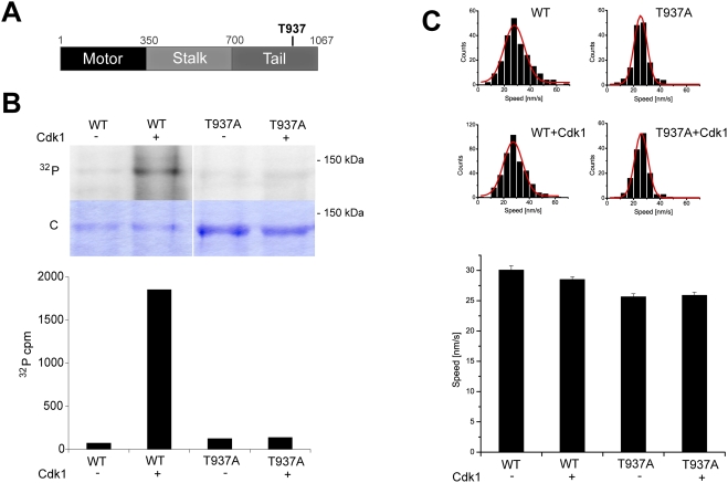
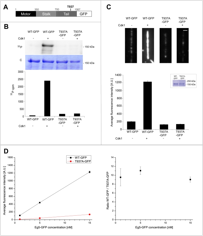
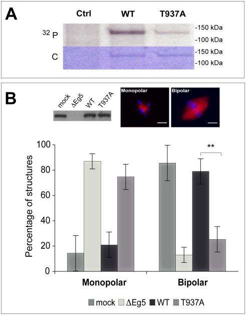
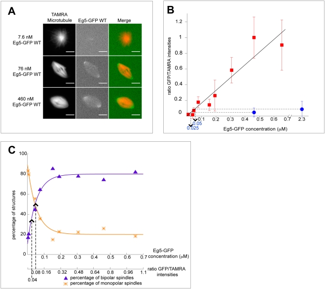
Methodology/principal findings: We investigate here the effect of these phosphorylations on kinesin-5 from Xenopus laevis, called Eg5. We find that phosphorylation at threonine 937 in the C-terminal tail of Eg5 by Cdk1 does not affect the velocity of Eg5, but strongly increases its binding to microtubules assembled in buffer. Likewise, this phosphorylation promotes binding of Eg5 to microtubules in Xenopus egg extract spindles. This enhancement of binding elevates the amount of Eg5 in spindles above a critical level required for bipolar spindle formation. We find furthermore that phosphorylation of Xenopus laevis Eg5 by Aurora A at serine 543 in the stalk is not required for spindle formation.

Conclusions/significance: These results show that phosphorylation of Eg5 by Cdk1 has a direct effect on the interaction of this motor with microtubules. In egg extract, phosphorylation of Eg5 by Cdk1 ensures that the amount of Eg5 in the spindle is above a level that is required for spindle formation. This enhanced targeting to the spindle appears therefore to be, at least in part, a direct consequence of the enhanced binding of Eg5 to microtubules upon phosphorylation by Cdk1. These findings advance our understanding of the regulation of this essential mitotic motor protein.


Conflict of interest statement

Competing Interests: The authors have declared that no competing interests exist.

Figures

Figure 1
Figure 1. Phosphorylation of Xenopus laevis Eg5 by Cdk1/cyclin B in vitro does not affect its velocity as measured in microtubule gliding assays.
(A) Schematic representation of the Eg5 sequence with the phosphorylation site for Cdk1 in the tail (threonine 937). (B) In vitro phosphorylation of wild-type Eg5 and Eg5T937A with [γ32P]ATP in buffer in the presence (+) or absence (−) of Cdk1/cyclin B (top). Autoradiography (32P) and coomassie-stained polyacrylamide gel (C) are shown. Quantitative measurement of radioactivity of the corresponding bands from the SDS-gel. The degree of phosphorylation for wild-type Eg5 is 87.9% and is strongly reduced to 4.0% for Eg5T937A. (C) Phosphorylation does not affect the velocity of Eg5 as measured in microtubule gliding assays: Histograms of gliding velocities of individual microtubules (top) and bar plot of averaged gliding velocities (bottom) produced by wild-type Eg5 (WT) and a non-phosphorylatable mutant (Eg5T937A) treated with or without Cdk1 kinase. Red lines are a Gauss fit, error bars are standard errors. The velocities of phosphorylated and unphosphorylated wild-type Eg5 (WT) are not statistically significantly different (p = 0.067, significance level 0.05); the 12% difference between the velocities of wild-type Eg5 (WT) and the non-phosphorylatable mutant (T937A) is statistically significant (p = 8.6×10⁻⁷, significance level 0.05).
Figure 2
Figure 2. Phosphorylation of Eg5 by Cdk1 increases Eg5 binding to microtubules in buffer.
(A) Schematic representation of the Eg5-GFP sequence. (B) In vitro phosphorylation of wild-type Eg5-GFP and Eg5T937A-GFP with [γ32P]ATP in buffer in the presence (+) or absence (−) of Cdk1/cyclin B. Autoradiography (32P) and coomassie-stained polyacrylamide gel (C) are shown. Quantitative measurement of radioactivity of the corresponding bands from the SDS-gel. The degree of phosphorylation of Eg5 wild-type is 75.6% and is strongly reduced to 5.9% for Eg5T937A-GFP. (C) Phosphorylation by Cdk1 increases the binding of Eg5 to microtubules in vitro: Representative examples for time-averaged images of the Eg5-GFP intensity on single immobilized microtubules as measured by TIRF microscopy (top). Wild-type Eg5-GFP (WT-GFP) and the non-phosphorylatable mutant (T937A-GFP) treated with ATP in the presence or absence of Cdk1/cyclin B were added to immobilized microtubules at a concentration of 15 nM. Scale bar is 1 µm. Bar plot of average Eg5-GFP signals (bottom) as obtained from 45–90 microtubules per condition as described above. Error bars are standard errors. Insert: Loading control showing the amount of Eg5-GFP in the phosphorylation reactions used in the experiment. (D) Average intensity signals (left) and intensity ratios (right) of Eg5-GFP and Eg5T937A-GFP on microtubules after Cdk1/cyclin B treatment plotted as a function of different Eg5 construct concentrations. For the average intensity signals (left), the values (in A.U.) are for WT and T937A at the concentration of 1 nM: 109±7 and 11±2, at the concentration of 5 nM: 439±17 and 40±3, at the concentration of 15 nM: 1226±39 and 136±8, respectively. Error bars are standard errors (in most cases obscured by the data symbol).
Figure 3
Figure 3. Spindles do not assemble in Eg5 depleted Xenopus egg extract in the presence of Eg5T937A.
(A) Phosphorylation of wild-type Eg5 and of Eg5T937A in mitotic Xenopus egg extract, showing a strong reduction of the degree of phosphorylation of Eg5T937A. As a control, an extract without recombinant Eg5 is shown (Ctrl). Autoradiography (32P) and Coomassie-stained polyacrylamide gel (C) are shown. (B) Western blot (top left) showing the amount of Eg5 in mock depleted extract (mock), in Eg5 depleted extract (ΔEg5) and in Eg5 depleted extract after addition of Eg5 wild-type (WT) or of Eg5T937A (T937A). Fluorescence images of a monopolar spindle after Eg5 depletion (top middle) and of a bipolar spindle after ‘mock’ depletion (top right). Graphs (bottom) representing the percentages of monopolar and bipolar spindles under the conditions as indicated. More than 120 structures per condition were included in the analysis. ** indicates statistical difference (p = 0.0031, significance level 0.05). Spindle assembly cannot be rescued by Eg5T937A at the concentration used. Microtubules are TAMRA-labeled (red) and DNA is stained with Hoechst (blue). Scale bar is 10 µm.
Figure 4
Figure 4. Cdk1 maintains the amount of Eg5 on spindle microtubules above a critical concentration required for spindle bipolarity.
(A) Fluorescence images of different representative structures at varying Eg5-GFP concentrations added to Eg5 depleted extract. Fluorescence of TAMRA-labeled microtubules (left) and Eg5-GFP (middle) is shown together with merged images (right). Scale bar is 10 µm. (B) Graph representing the ratios of averaged GFP intensities per TAMRA intensities as a function of varying wild-type Eg5-GFP concentrations (red squares) and mutated Eg5T937A-GFP (blue circle). The ratios were normalized setting the ratio at 0.46 µM Eg5-GFP to 1. The line is a linear fit to the wild-type data (f(x) = 1.62 µM−1*x). Error bars indicate the standard error. (C) Graph illustrating the percentages of bipolar spindles (purple triangles) and of monopolar spindles (orange stars) as a function of the Eg5-GFP concentration. Depending on the condition, between 9 and 76 (on average 28) structures were counted per data point. The lines are fits to the data with using f(x) = 80%−69%*exp(−c/0.054 µM) for percentages of bipolar spindles and f(x) = 20%+69%*exp(−c/0.054 µM) for percentages of monopolar spindles. Concentrations on the x-axis were transformed into fluorescence ratios using the regression from B. The dashed lines indicate the fluorescence ratios for mutated Eg5T937A-GFP at the concentrations used in B.

References

    1. Murray AW. Recycling the cell cycle: cyclins revisited. Cell. 2004;116:221–234. - PubMed
    1. Gruss OJ, Vernos I. The mechanism of spindle assembly: functions of Ran and its target TPX2. J Cell Biol. 2004;166:949–955. - PMC - PubMed
    1. Malumbres M, Barbacid M. Cell cycle kinases in cancer. Curr Opin Genet Dev. 2007;17:60–65. - PubMed
    1. Hildebrandt ER, Hoyt MA. Mitotic motors in Saccharomyces cerevisiae. Biochim Biophys Acta. 2000;1496:99–116. - PubMed
    1. Sharp DJ, Rogers GC, Scholey JM. Microtubule motors in mitosis. Nature. 2000;407:41–47. - PubMed

Publication types

LinkOut - more resources