. 2008;3(12):e3942.
doi: 10.1371/journal.pone.0003942. Epub 2008 Dec 16.

Heterosubtypic neutralizing monoclonal antibodies cross-protective against H5N1 and H1N1 recovered from human IgM+ memory B cells

Affiliations

Heterosubtypic neutralizing monoclonal antibodies cross-protective against H5N1 and H1N1 recovered from human IgM+ memory B cells

Mark Throsby et al. PLoS One. 2008.

Abstract

Background: The hemagglutinin (HA) glycoprotein is the principal target of protective humoral immune responses to influenza virus infections but such antibody responses only provide efficient protection against a narrow spectrum of HA antigenic variants within a given virus subtype. Avian influenza viruses such as H5N1 are currently panzootic and pose a pandemic threat. These viruses are antigenically diverse and protective strategies need to cross protect against diverse viral clades. Furthermore, there are 16 different HA subtypes and no certainty the next pandemic will be caused by an H5 subtype, thus it is important to develop prophylactic and therapeutic interventions that provide heterosubtypic protection.

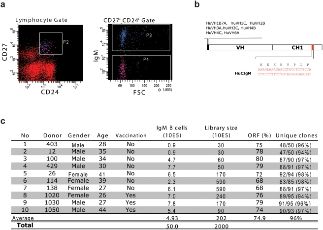
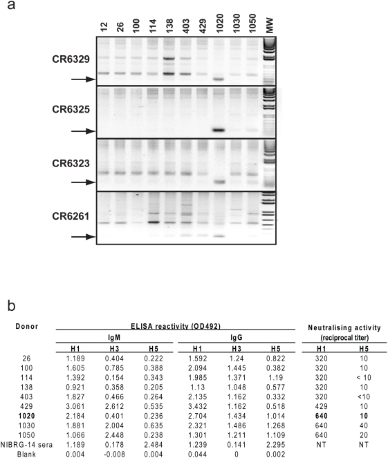
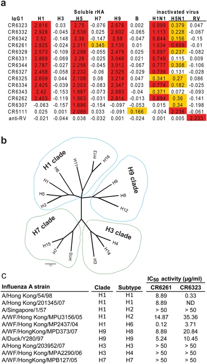
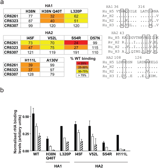
Methods and findings: Here we describe a panel of 13 monoclonal antibodies (mAbs) recovered from combinatorial display libraries that were constructed from human IgM(+) memory B cells of recent (seasonal) influenza vaccinees. The mAbs have broad heterosubtypic neutralizing activity against antigenically diverse H1, H2, H5, H6, H8 and H9 influenza subtypes. Restriction to variable heavy chain gene IGHV1-69 in the high affinity mAb panel was associated with binding to a conserved hydrophobic pocket in the stem domain of HA. The most potent antibody (CR6261) was protective in mice when given before and after lethal H5N1 or H1N1 challenge.

Conclusions: The human monoclonal CR6261 described in this study could be developed for use as a broad spectrum agent for prophylaxis or treatment of human or avian influenza infections without prior strain characterization. Moreover, the CR6261 epitope could be applied in targeted vaccine strategies or in the design of novel antivirals. Finally our approach of screening the IgM(+) memory repertoire could be applied to identify conserved and functionally relevant targets on other rapidly evolving pathogens.


Conflict of interest statement

Competing Interests: MT., EvdB, MJ, AB, FC, ED, JtM., JdK. and JG are employees of Crucell Holland BV. PA and IL are employees of Algonomics NV.

Figures

Figure 1
Figure 1. Construction of IgM+ memory B cell libraries.
a) Donor lymphocytes were isolated by Ficoll-plaque from heparinized blood and stained for the phenotypic markers CD27, CD24 and IgM. CD24+ CD27+ cells were gated and the IgM+ cells within this gate sorted directly into Trizol for RNA extraction. b) RT-PCR was performed using a pool of 5′ oligonucleotide primers that cover all VH gene families and a 3′ oligonucleotide primer that anneals in a region of the CH1 domain of Cμ distinct from other immunoglobulin isotypes. c) Using cDNA generated in this way, 10 individual scFv libraries were constructed as described previously . Donors 1020, 1030 and 1050 had been vaccinated with the Dutch 2005 seasonal influenza vaccine 7 days prior to collection of blood. All libraries demonstrated a high percentage of correct scFv ORF's and diversity based on unique HCDR3 sequence.
Figure 2
Figure 2. Sequence analysis of VH genes from selected anti-H5 HA scFv.
(a) Alignment of VH amino acid sequences from scFv containing the same V(D)J rearrangement as CR6323 or CR6261 with the germline IGHV1-69. Note that although multiple mutations are present in the CDR1 and CDR2 loops Phe at position 54 is conserved (Kabat numbering). scFv that were neutralizing in an IgG1 format are shown in bold (b) DNA alignment and amino acids for the HCDR2 of (b) CR6323 and (c) CR6261 with related mAbs. Note the identical codon usage and conservation of silent mutations suggesting a clonal origin.
Figure 3
Figure 3. Cross-clade H5N1 neutralizing activity and HA subunit localization.
(a) Neutralizing activity of purified IgG1 against 100 TCID50 of H5N1 viruses, the 50% neutralizing concentration (IC50) was calculated by the Spearman-Karber method. (b) Immunoblot of H5 rHA probed with indicated IgG1. Molecular weight marker (MW) and subunit locations indicated.
Figure 4
Figure 4. PCR screen of individual donor libraries for neutralizing mAbs and donor serology.
(a) PCR amplification of cDNA from each donor IgM+ memory B cell library using oligonucleotide pairs designed so their 3′ ends specifically anneal in the HCDR1 and HCDR3 regions. Donors are indicated at the top of the figure. The expected size of the amplified fragment is indicated with an arrow. The identity of the bands was confirmed by sequencing (b) Binding and neutralizing activity of donor serum collected at the same time as the B cells used for library construction (note serum was not available for donor 12). IgM and IgG ELISA reactivity was measured against rHA and neutralizing activity against H1N1 (A/Hong Kong/54/98) and H5N1 (A/Vietnam/1203/04). Donor 1020 who was PCR positive for the tested neutralizing mAbs is indicated in bold.
Figure 5
Figure 5. Heterosubtypic binding and neutralizing activity.
(a) The binding activity of the IgG1 panel (5 µg/ml) was measured against 0.5 µg/ml directly coated recombinant HA antigen (see methods for strain designation), BPL inactivated NIBRG-14 (HA from A/Vietnam/1194/04) or BPL inactivated H1N1 (A/New Caledonia/1/99). Detection was performed with mouse anti-human IgG-HRP and results given as OD492 nm. Control antigens were Influenza B virus (B/Ohio/1/05) and Rabies vaccine (RIVM, Bilthoven). Anti-rabies virus (RV) IgG1 (IGHV1-69) was used as a negative control. ELISA values>10× background or between 10× and 3× background are coloured red and yellow respectively. (b) Phylogenetic tree of amino acid sequences at the subtype level, division of subtypes by group is indicated by coloured balloons (blue H1 and H9, green H3 and H7). (c) Neutralizing activity of purified IgG1 against 100 TCID50 of influenza A viruses, IC50 calculated as in Fig. 3.
Figure 6
Figure 6. Identification of the antigenic region of neutralizing mAbs.
(a) FACS binding of IgG1 to surface expressed H5 rHA was measured after sequential treatment with trypsin (solid bars), pH 4.9 buffered medium (open bars) and DTT (striped bars) and expressed as percentage binding to untreated rHA from two independent experiments (mean±s.e.m.). (b) Global view of CR6261, VL is rose, VH is blue, HCDR2 is green, Thr56 red and Phe54 green. (c) Surface representation of trimeric H1 HA (A/South Carolina/1/18; 1RUZ). HA1 subunits are green, burgundy and pink, HA2 subunits yellow, blue and orange. The hydrophobic pocket is white and shown magnified as an inset with the HCDR2 of CR6261 in green docked to the structure. HA residues around and forming the hydrophobic pocket are labelled in black and HCDR2 residues in the pocket are green. (d) perpendicular view of the hydrophobic pocket down the axis of helix 38–55 occupied by Phe54 of HCDR2 in purple.
Figure 7
Figure 7. The hydrophobic pocket region in the stem domain of different HA subtypes.
Surface representation of the hydrophobic pocket recognized by CR6261 from crystal structures of H5 (2IBX) and H9 (1JSD) and the corresponding region from crystal structures of H3 (1MQM) and H7 (1TI8). HA residues around and forming the hydrophobic pocket are indicated in black, H3 numbering is used throughout for consistency. Note the H38N and Q40T replacements in the H3 and H7 structures that introduce a potential glycosylation site in the region.
Figure 8
Figure 8. Generation of CR6261 neutralisation escape variants.
(a) The hydrogen bond interaction of HA2 His111 with HA1 Thr318 is shown (circled red) in the H5 structure 2IBX. (b) An H3 structure (1MQM) is shown where the peptidic plane re-orientation of Thr318 is indicated by a red arrow.
Figure 9
Figure 9. Binding of CR6261 to H5 HA point mutants.
(a) FACS binding of IgG1 to surface expressed mutants of H5 rHA. Mean fluorescent intensity (MFI) values were normalised by NIBRG-14 antiserum reactivity to the corresponding mutant and expressed as the average percentage of wild type H5 rHA binding from two independent experiments (see key for colour code). Adjacent are sequence alignments of the corresponding region, mutated residues are boxed. (b) FACS binding activity to WT or mutant H5 rHA performed as above for CR6261 (solid bars), CR6261 with the HCDR2 mutation F54L (open bars) or F54A (striped bars). MFI was normalised as above and expressed as mean±s.e.m. arbitrary units from two independent experiments.
Figure 10
Figure 10. In vivo protective activity of CR6261 against wild type H5N1 and H1N1 strains.
Kaplan-Meier survival curves of BALB/c mice were injected (i.p.) with CR6261 or irrelevant control (15 mg/kg) then challenged 24 h later (i.n.) with (a) 10 LD50 of A/Vietnam/1203/04 (n = 5) or (b) 25 LD50 A/WSN/33 (n = 10) and observed daily for a period of 21 days. Haematoxylin-Eosin stained lung sections taken 6 d.p.i from (c) CR6261 or (d) control mAb treated mice (1 d.p.i.) and challenged with 25 LD50 A/Hong Kong/156/97. (e) Survival (upper panel), mean±s.e.m. body weight, (middle panel) and median clinical signs (bottom panel) of mice (n = 10) challenged as in (c,d) and injected i.v. with 15 mg/kg CR6261 IgG1 3 (dark blue), 4 (light blue), 5 (green) or 6 days (orange) or control mAb (black dotted line) 4 days after challenge (see colored arrows in bottom panel). The day five time point at which therapeutic efficacy is lost is indicated by a grey box.

References

    1. Peiris JSM, de Jong MD, Guan Y. Avian Influenza Virus (H5N1): a Threat to Human Health. Clin Microbiol Rev. 2007;20:243–267. - PMC - PubMed
    1. WHO. Cumulative Number of Confirmed Human Cases of Avian Influenza A/(H5N1) Reported to WHO. 2008 http://www.who.int/csr/disease/avian_influenza/country/cases_table_2008_....
    1. Chin PS, Hoffmann E, Webby R, Webster RG, Guan Y, et al. Molecular evolution of H6 influenza viruses from poultry in Southeastern China: prevalence of H6N1 influenza viruses possessing seven A/Hong Kong/156/97 (H5N1)-like genes in poultry. J Virol. 2002;76:507–516. - PMC - PubMed
    1. Peiris M, Yuen KY, Leung CW, Chan KH, Ip PL, et al. Human infection with influenza H9N2. Lancet. 1999;354:916–917. - PubMed
    1. Ma W, Vincent AL, Gramer MR, Brockwell CB, Lager KM, et al. Identification of H2N3 influenza A viruses from swine in the United States. Proc Natl Acad Sci U S A. 2007;104:20949–20954. - PMC - PubMed

Publication types

MeSH terms