. 2008;3(12):e3949.
doi: 10.1371/journal.pone.0003949. Epub 2008 Dec 16.

BRAF activation initiates but does not maintain invasive prostate adenocarcinoma

Affiliations

BRAF activation initiates but does not maintain invasive prostate adenocarcinoma

Joseph H Jeong et al. PLoS One. 2008.

Abstract

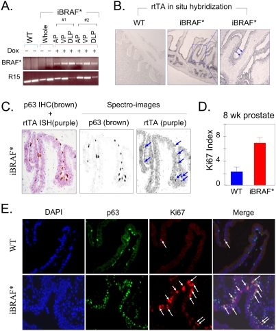
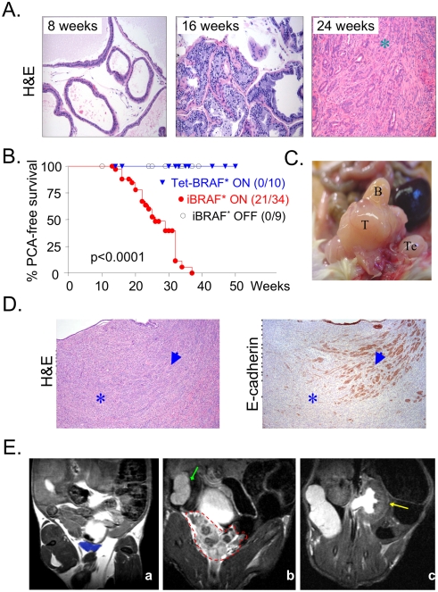
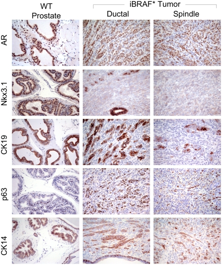
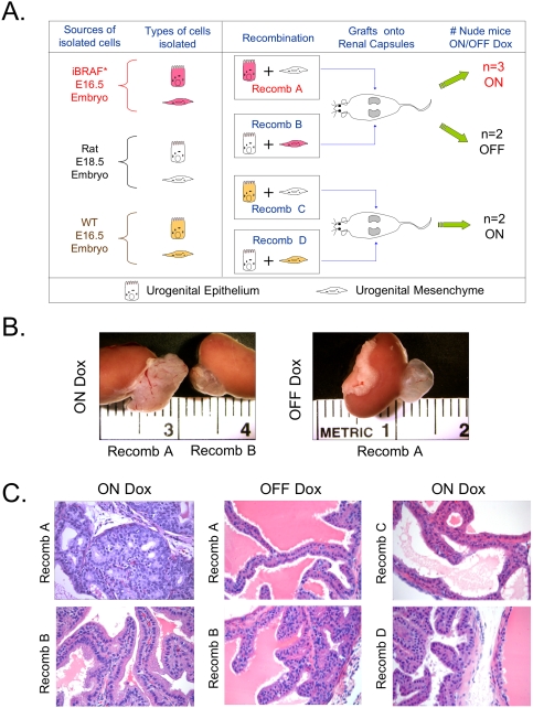
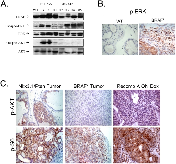
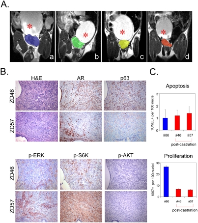
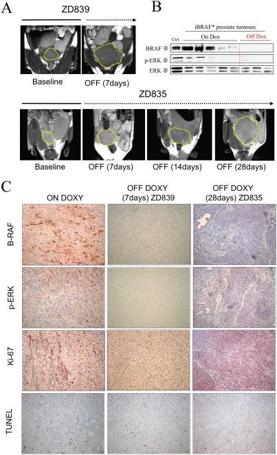
Prostate cancer is the second leading cause of cancer-related deaths in men. Activation of MAP kinase signaling pathway has been implicated in advanced and androgen-independent prostate cancers, although formal genetic proof has been lacking. In the course of modeling malignant melanoma in a tyrosinase promoter transgenic system, we developed a genetically-engineered mouse (GEM) model of invasive prostate cancers, whereby an activating mutation of BRAF(V600E)--a mutation found in approximately 10% of human prostate tumors--was targeted to the epithelial compartment of the prostate gland on the background of Ink4a/Arf deficiency. These GEM mice developed prostate gland hyperplasia with progression to rapidly growing invasive adenocarcinoma without evidence of AKT activation, providing genetic proof that activation of MAP kinase signaling is sufficient to drive prostate tumorigenesis. Importantly, genetic extinction of BRAF(V600E) in established prostate tumors did not lead to tumor regression, indicating that while sufficient to initiate development of invasive prostate adenocarcinoma, BRAF(V600E) is not required for its maintenance.


Conflict of interest statement

Competing Interests: The authors have declared that no competing interests exist.

Figures

Figure 1
Figure 1. Transgene BRAF* expression in prostate epithelium drives aberrant proliferation of the p63+ prostatic basal cells.
A. Transgene BRAF* transcript was detected in all three lobes (AP, anterior prostate; VP, ventral prostate; DLP, dorsolateral prostate) of the prostate glands from two independent 8-week-old bi-transgenic iBRAF* male mice on doxycycline by transgene-specific RT-PCR. As controls, whole prostate glands were isolated from WT or iBRAF* off doxycycline mice (n = 2 for each). Ribosomal protein R15 was used as an internal control for RT-PCR. B. RNA in situ hybridization (RISH) using rtTA riboprobe documented expression of BRAF* transgene in prostate epithelial gland (200×). Arrows indicate the expression of rtTA in luminal cell compartment in zoom-in with higher magnification (400×). C. Expression of BRAF* transgene was detected in both luminal cells and p63+ basal cells of the prostate epithelium of 8-week-old bi-transgenic iBRAF* male mice on doxycycline using dual serial staining of p63 IHC and rtTA RISH. After quick incubation of slides with antibody against p63 for 10 minutes with IHC procedure, RISH procedure with rtTA RNA probe was followed. Brown color for p63+ basal cells by IHC and purple color for rtTA expression by RISH were differentially detected by spectro-imaging machine. Note the co-localization of strong rtTA expression and p63 immuno-reactivity in approximately 50% of p63+ basal cells (arrows). D. Ki67 staining of histologically-normal prostate glands in 8-week-old males showed increased proliferation index in iBRAF* transgenic (on doxycycline) compared to WT. E. Co-immunofluorescence study in prostate glands isolated from 8-week-old WT and iBRAF* transgenic (on doxycycline) males showed the expansion of p63+ basal cells in the prostate glands of iBRAF* mice. Approximately half of the p63+ cells were in proliferation as measured by co-staining with Ki67 (see arrows).
Figure 2
Figure 2. iBRAF* transgenic males develop invasive adenocarcinoma of the prostate.
A. Time-course histological analysis of prostate samples from iBRAF* transgenic mice (on doxycycline) at the indicated ages. Hyperplastic lesions were detected in a 16-week-old iBRAF* transgenic mouse on doxycycline. Up to an age of 24 weeks, approximately 50% of iBRAF* transgenic mice developed PCA. B. Kaplan-Meier analysis of PCA-free survival in iBRAF* (on doxycycline, n = 34), iBRAF* (off doxycycline, n = 9), and Tet-BRAF* (on doxycycline, n = 10) transgenic males. Only double transgenic iBRAF* mice given doxycycline in their drinking water developed PCA with an average latency of 24 weeks (standard deviation = ±6 weeks). C. Gross morphology of a prostate tumor from an iBRAF* transgenic mouse. T indicates tumor; B, bladder; and Te, testis. D. The transition from ductal (arrowhead) to spindle (asterisk) morphology is coincident with loss of E-cadherin expression in iBRAF* PCA, consistent with EMT (epithelial-mesenchymal transition). E. Detection of PCA in live mice by MRI. Panel a is a representative image of a WT mouse with normal prostate (highlighted in blue). Panel b is a representative MR image of PCA in an iBRAF* male on doxycycline (red outline; note heterogeneous signals within the lesion). Panel c is a representative MR image of hydronephrosis detected in an iBRAF* mouse with PCA. Note enlarged distended kidney (yellow arrow) in contrast to normal kidney in panel b (green arrow), indicating accumulation of urine in the kidney due to outflow obstruction by the tumor.
Figure 3
Figure 3. iBRAF* prostate tumors exhibit epithelial lineage markers.
Both ductal and spindled components of iBRAF* prostate tumors express epithelial lineage markers: CK14 and p63 for basal cell, AR and Nkx3.1 for luminal cell, and CK19 for transit-amplifying cells.
Figure 4
Figure 4. iBRAF* embryonic urogenital epithelium drives basaloid hyperplasia in the tissue recombination system.
A. Schematic representation of the tissue recombination protocol. Recombinants of iBRAF* transgenic mouse epithelium and rat mesenchyme (Recomb A) were grafted under the right kidney capsules of adult nude mice. The converse recombinants (iBRAF* transgenic mouse mesenchyme and rat epithelium; Recomb B) were also transplanted in the same manner into the left kidney capsule of the same mice. Three of the grafted mice were fed doxycycline drinking water, and two were not. As controls, recombinants of non-transgenic wild-type mouse epithelium and rat mesenchyme (Recomb C) and the converse recombinants (Recomb D) were grafted in the same manner. Grafts were harvested 6 weeks later. B. Macroscopic images show that grafts with Recomb A on doxycycline are larger than both grafts with Recomb B on doxycycline and grafts with Recomb A off doxycycline. C. Histological analysis of these tissue grafts confirmed basaloid hyperplasia only in grafts with Recomb A on doxycycline, while others showed normal histology.
Figure 5
Figure 5. iBRAF* prostate tumors are AKT-independent.
A, B. p-ERK activation in iBRAF* PCA tumors (n = 5; on doxycycline) was detected by immunoblotting analysis (A) and IHC (B). In contrast, p-AKT activation was not detected in all iBRAF* PCA tumors (n = 5; on doxycycline). Controls (a & b) were two prostate cancer samples from Pten-null mice and WT was prostate glands from a WT mouse. C. p-AKT immunoreactivity was not detected on iBRAF* PCA tumors (n = 17) and Recomb A (a recombinant of iBRAF* transgenic mouse epithelium and rat mesenchyme on doxycycline) by IHC, but strong immunoreactivity of p-S6 was still present. PCA sample from Nkx3.1−/−, Pten+/− compound mutant mice were used as control.
Figure 6
Figure 6. iBRAF* PCA progress to indolent androgen-independence after castration.
A. The reduction of tumor volumes was monitored by MRI over a 4-week time course after castration (a, tumor (blue) 1 week after castration; b, tumor (green) 2 weeks after castration; c, tumor (yellow) 3 weeks after castration; d, tumor (orange) 4 weeks after castration). Asterisk indicates bladder. B. Histological examination confirmed the presence of prostatic tumor cells in two castrated iBRAF* mice (#46 and #57). The two post-castration tumors were positive for AR (a prostatic luminal cell marker) and p63 (a basal cell marker) by IHC. Also, the post-castration tumor cells were negative for p-AKT, but remained strongly positive for p-ERK and p-S6K on IHC. C. The post-castration tumor cells were viable and growing, not residual tumor remnants, as manifested by low apoptosis index (1.2 (+/−0.45) and 1.4 (+/−0.55) per 100 nuclei for #46 and #57, respectively) on TUNEL, comparable to pre-castration tumor (1.0 (+/−0.71)) (top), and active proliferation by Ki67 staining, albeit at rate much lower than that of pre-castration tumor (6.8 (+/−3.6) and 6.2 (+/−2.2) for #46 and #57, respectively and 26.6 (+/−3.6) in the pre-castrated tumors) (bottom).
Figure 7
Figure 7. iBRAF* prostate tumors do not require BRAF activation for their tumor maintenance.
A. Tumor size change was monitored by serial MRI imaging before (baseline; solid line) and after (indicated periods; dotted line) doxycycline withdrawal (7 days for ZD839 and 28 days for ZD835). Yellow line indicates tumor boundary. B. B-RAF expression and p-ERK activation were not detected by immunoblotting analysis in most of off doxycycline iBRAF* PCA tumors (n = 3), although one of the off doxycycline iBRAF* tumor samples (ZD835) showed weak p-ERK activation. C. Immunohistological examination using antibodies against B-RAF and p-ERK confirmed the repression of B-RAF* transgene repression during off doxycycline periods (7 days for ZD839 and 28 days for ZD835). However, the tumors were viable and still growing, as manifested by low apoptosis on TUNEL and active proliferation by Ki67 staining.

References

    1. Ellwood-Yen K, Graeber TG, Wongvipat J, Iruela-Arispe ML, Zhang J, et al. Myc-driven murine prostate cancer shares molecular features with human prostate tumors. Cancer Cell. 2003;4:223–238. - PubMed
    1. Hill R, Song Y, Cardiff RD, Van Dyke T. Heterogeneous tumor evolution initiated by loss of pRb function in a preclinical prostate cancer model. Cancer Res. 2005;65:10243–10254. - PubMed
    1. Kim MJ, Bhatia-Gaur R, Banach-Petrosky WA, Desai N, Wang Y, et al. Nkx3.1 mutant mice recapitulate early stages of prostate carcinogenesis. Cancer Res. 2002;62:2999–3004. - PubMed
    1. Trotman LC, Niki M, Dotan ZA, Koutcher JA, Di Cristofano A, et al. Pten dose dictates cancer progression in the prostate. PLoS Biol. 2003;1:E59. - PMC - PubMed
    1. Wang L, Cunningham JM, Winters JL, Guenther JC, French AJ, et al. BRAF mutations in colon cancer are not likely attributable to defective DNA mismatch repair. Cancer Res. 2003;63:5209–5212. - PubMed

Publication types

MeSH terms