. 2008;3(12):e3894.
doi: 10.1371/journal.pone.0003894. Epub 2008 Dec 16.

Oncologic trogocytosis of an original stromal cells induces chemoresistance of ovarian tumours

Affiliations

Oncologic trogocytosis of an original stromal cells induces chemoresistance of ovarian tumours

Arash Rafii et al. PLoS One. 2008.

Abstract

Background: The microenvironment plays a major role in the onset and progression of metastasis. Epithelial ovarian cancer (EOC) tends to metastasize to the peritoneal cavity where interactions within the microenvironment might lead to chemoresistance. Mesothelial cells are important actors of the peritoneal homeostasis; we determined their role in the acquisition of chemoresistance of ovarian tumours.

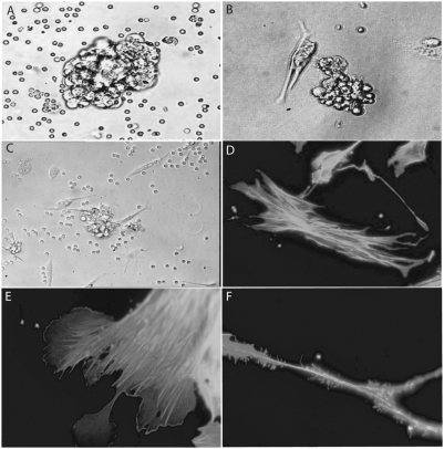
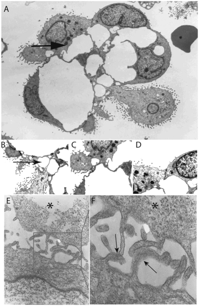
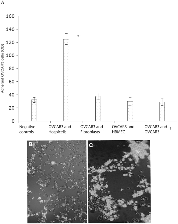
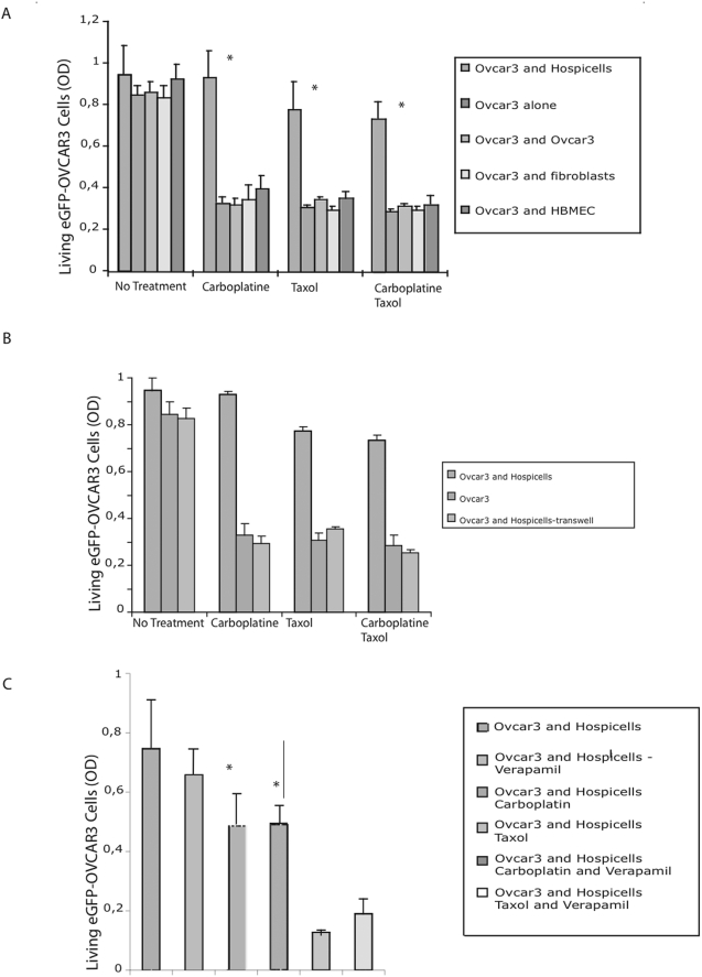
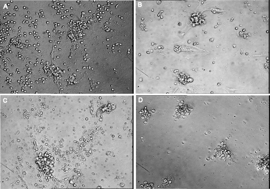
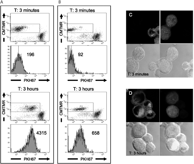
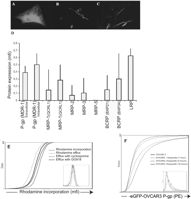
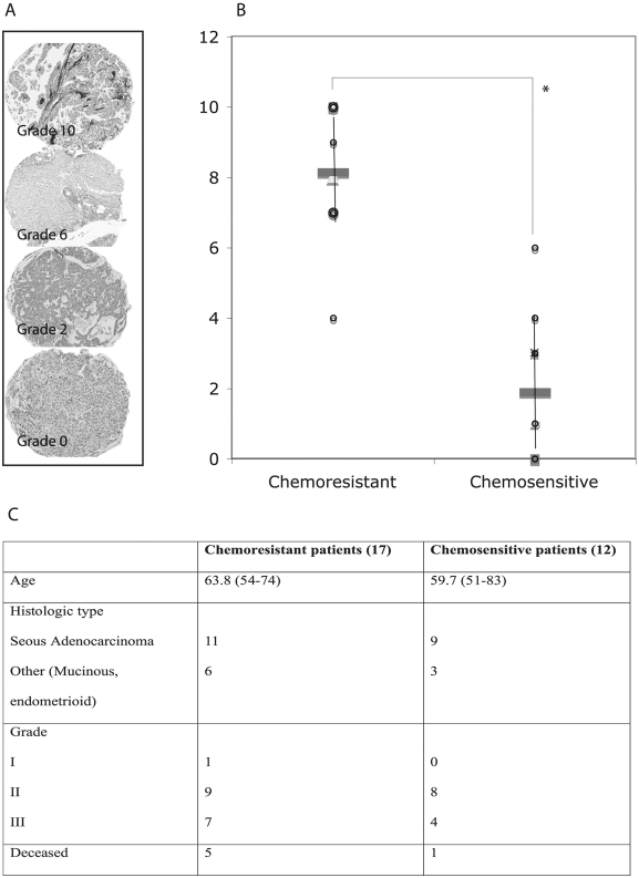
Methodology/principal findings: We isolated an original type of stromal cells, referred to as "Hospicells" from ascitis of patients with ovarian carcinosis using limiting dilution. We studied their ability to confer chemoresistance through heterocellular interactions. These stromal cells displayed a new phenotype with positive immunostaining for CD9, CD10, CD29, CD146, CD166 and Multi drug resistance protein. They preferentially interacted with epithelial ovarian cancer cells. This interaction induced chemoresistance to platin and taxans with the implication of multi-drug resistance proteins. This contact enabled EOC cells to capture patches of the Hospicells membrane through oncologic trogocytosis, therefore acquiring their functional P-gp proteins and thus developing chemoresistance. Presence of Hospicells on ovarian cancer tissue micro-array from patients with neo-adjuvant chemotherapy was also significantly associated to chemoresistance.

Conclusions/significance: This is the first report of trogocytosis occurring between a cancer cell and an original type of stromal cell. This interaction induced autonomous acquisition of chemoresistance. The presence of stromal cells within patient's tumour might be predictive of chemoresistance. The specific interaction between cancer cells and stromal cells might be targeted during chemotherapy.


Conflict of interest statement

Competing Interests: The authors have declared that no competing interests exist.

Figures

Figure 1
Figure 1. Isolation of Hospicells.
A. Cellular aggregates from ascitic fluid of patients with ovarian carcinosis in suspension (×40). B. Cellular aggregates on culture plates (×40). C. Gradual disaggregation of strongly interacting stromal cells (“Hospicells”) (thick arrow) and cancer cells (thin arrow) at day 10 days of culture (×20). D. Fluorescence microscopy (actin immunostaining) showing the “hammock-like” active cytoskeleton of freshly isolated Hospicells (×60). E. Lamellipodal formation at the extremity of the Hospicells containing active actin fibers. F. Details of a Hospicell's fillopode demonstrating numerous “comb-like filaments”.
Figure 2
Figure 2. Electronic microscopy analysis of ovarian cancer aggregates and primocultures of Hospicells and OVCAR3.
A. Electronic microscopy sections showing EOC cell and Hospicells aggregates displaying numerous interactions between cancer cells (thin arrow) and Hospicells (thick arrow and (*)). B, C and D. Details of different interactions: through thin pseudopods (B and C), or through large membranous contact (D). Hospicells are marked with an (*) sign. E. Co-cultures of freshly isolated Hospicells (*) and OVCAR3 displaying the network of Hospicells' pseudopods enhancing contact with EOC cells. F. Cell membrane fusion (arrows) between Hospicells (*) and cancer cells pseudopods in co-cultures.
Figure 3
Figure 3. Specific interaction between OVCAR3 cells and Hospicells.
A. Adhesion of eGFP-OVCAR3 to Hospicells compared to that of fibroblasts and HBMEC (Human Bone Marrow Endothelial Cells). B and C. Hospicells were seeded on Matrigel coated culture plates allowing them to form a network. eGFP-OVCAR3 cells were then added. eGFP-OVCAR3 cells developed on the Hospicells network.
Figure 4
Figure 4. Chemoresistance induced by Hospicells.
A. Induced chemoresistance when Hospicells co-cultured with OVCAR3 cells over-expressing e-GFP are treated with 22.2 µM carboplatin and/or 1.4 µM paclitaxel (* p>0.05). Fibroblasts, HBMEC and OVCAR3 cells were used as controls. B. Same assay using a transwell co-culture system. C. Reversal of chemoresistance by treating the co-culture by 50 µM verapamil. Cell density is expressed as the optical density (OD), means with standard deviations (6 replicates were used per experiments. The experiments were performed 3 times).
Figure 5
Figure 5. Effect of chemotherapy on freshly isolated ECOA.
ECOA were isolated and cultured on RPMI media supplemented by calf fetal serum (10%) and antibiotics. Carboplatin and paclitaxel treatment were performed using the same conditions as in vitro. The effect of chemotherapy was assessed by conventional microscopy. A and C. ECOA freshly isolated in culture before treatment. B. Day 3 after treatment by carboplatin (22.2 µM). D. Day 3 after treatment by paclitaxel (1.4 µM).
Figure 6
Figure 6. Intercellular transfer or “oncologic trogocytosis”.
A and B. Flow cytometry of OVCAR3 cells or Hospicells co-cultured with Cell Tracker™ Orange CMTMR-labelled cells for 3 minutes or 3 hours. A. The Hospicells were labelled with the membranous lipophilic dye PKH67B. The OVCAR3 cells were labelled with PKH67. The value given in the plot is the mean PKH67 fluorescence intensity. C. Confocal microscopy of PKH67-stained hospicells (green) and CMTMR-labelled OVCAR3 cells (red) after 3 minutes incubation. D. Uptake of PKH67 by OVCAR3 cells is observed at 3 hours indicating membrane transfer without cytoplasmic transfer.
Figure 7
Figure 7. Expression and functionality of MDR proteins in hospicells.
A, B and C. Immunofluorescence staining of P- Glycoprotein (P-gp) (A), Lung Resistance Protein (LRP) (B), and Breast Cancer Resistance Protein (BCRP) (C) in Hospicells isolated from ascitic fluid from 5 patients (Multidrug Resistance protein (MRP)). D. Expression of MDR proteins (mean fluorescence intensity (mfi)) assessed by FACS (mfi (+SD)). E. Flow cytometry of functionality of P-gp1 expressed by Hospicells: control, e.g. rhodamine incorporation (green curve – mfi = 22), rhodamine excretion (dashed curve - mfi = 35); inhibition of excretion by cyclosporine (red curve - mfi = 25) and GG918 (blue curve- mfi 26). F. Intercellular transfer of labelled P-gp on co-culture of hospicells and OVCAR3 cells for 1 hour, 5 hours and 18 hours.
Figure 8
Figure 8. Clinical relevance of the presence of Hospicells among ovarian cancer tumors.
Tissue Micro-arrays were prepared from tumors originating from 29 patients who underwent a neo-adjuvant chemotherapy. A. Different densities of Hospicells as represented by CD10 staining. Morphological controls were used to rule out the staining of endothelial cells. B. Samples from chemoresistant patients had significantly higher density of Hospicells compared to chemosensitive patients (* p<0.05). Three different spots were used to characterize the Hospicells' density for each patient.

Similar articles

Cited by

References

    1. Jemal A, Siegel R, Ward E, Hao Y, Xu J, et al. Cancer statistics. CA Cancer J Clin. 2008;58:71–96. - PubMed
    1. Bhoola S, Hoskins WJ. Diagnosis and management of epithelial ovarian cancer. Obstet Gynecol. 2006;107:1399–1410. - PubMed
    1. Eisenkop SM, Spirtos NM, Lin WC. “Optimal” cytoreduction for advanced epithelial ovarian cancer: a commentary. Gynecol Oncol. 2006;103:329–35. - PubMed
    1. Pfisterer J, Ledermann JA. Management of platinum-sensitive recurrent ovarian cancer. Semin Oncol. 2006;33:S12–6. - PubMed
    1. Di Nicolantonio F, Mercer SJ, Knight LA, Gabriel FG, Whitehouse PA, et al. Cancer cell adaptation to chemotherapy. BMC Cancer. 2005;5:78. - PMC - PubMed

MeSH terms

Substances