. 2009;2(3):286-99.
Epub 2008 Nov 6.

The laboratory opossum (Monodelphis domestica) as a natural mammalian model for human cancer research

Affiliations

The laboratory opossum (Monodelphis domestica) as a natural mammalian model for human cancer research

Zhiqiang Wang et al. Int J Clin Exp Pathol. 2009.

Abstract

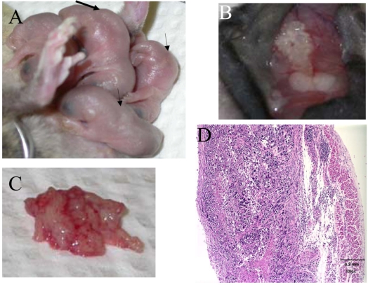
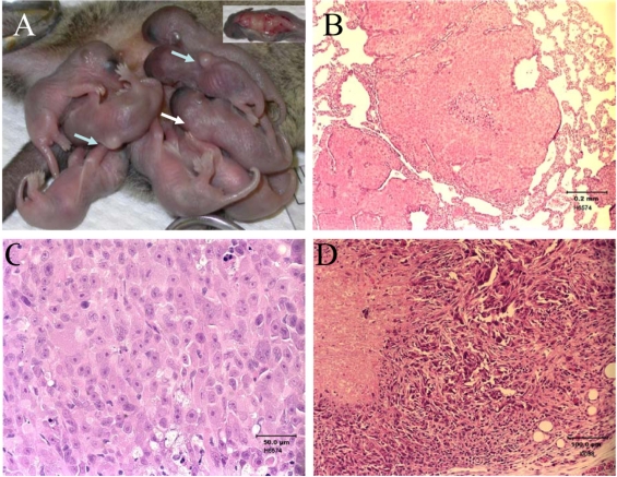
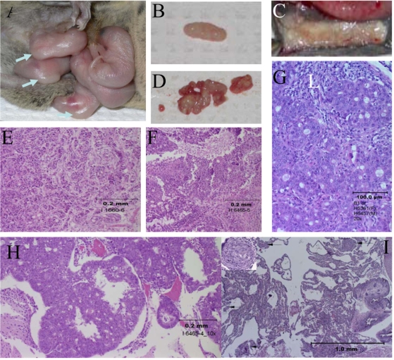
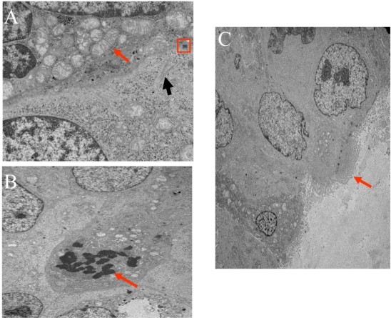
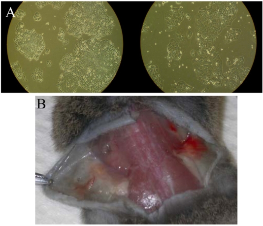
This study established that human cancer cells (A375 melanoma, HT-29 colon cancer, PC-3p prostate cancer) that were xenografted into suckling opossums could proliferate and globally metastasize as early as 11 days after injection. Light and electron microscopic examinations (HT-29 colon cancer) determined that the cellular features exhibited by the xenogeneic human tumors grown in laboratory opossums were consistent with those observed in tumors removed from humans. The tumor induction rate, patterns of tumor growth and regression, and types of host immune responses against the xenografted tumors were influenced by injection dosages, injection sites and injection ages of suckling opossums. The results highlight the value of the opossum model as a natural in vivo system for investigating human cancer growth, metastasis and apoptosis at the cellular and molecular levels; enhancing identification of tumor associated antigens or T cell epitopes through use of humoral and cellular expression cloning techniques; elucidating mechanisms utilized by tumor cells to evade host immunosurveillance; and devising diagnostic and therapeutic methods for cancer treatment.

Keywords: Monodelphis domestica; Opossum; animal model; human cancer.


Figures

Figure 1
Figure 1
Human melanoma cells xenografted into suckling young opossums. A. Suckling pups at two weeks of age (litter 6, Table 1A). Amelanocytic tumors, as pointed by arrows, were induced by s.c. injection of 0.25 × 10⁶ A375 cells at the age of 2 days. B. Regressing xenografted human melanoma at week 5 after injection (litter 6). Whitish dots and streaks were evident in the tumors. C. A large solid tumor located in the muscular tissues of chest wall in a pup of litter 6. This 5-w.o. tumor appeared to have been newly established by comparison to the regressing s.c. tumor shown in B. D. Tumor of 4-w.o. Monodelphis (litter 6). Proliferation of tumor cells was associated with considerable cell death and mineralization. Minimal host inflammatory cells were noted. Hematoxylin and eosin (HE), bar = 0.2 mm.
Figure 2
Figure 2
Human prostate cancer cells xenografted into suckling opossums. A. Suckling opossums at 3 weeks old (litter 1, Table 1B). The tumors (arrows) were induced by s.c. injection of 0.25 × 10⁶ PC-3p cells on the day of birth (0-d.o.). During week 4, one pup of litter 1 (white arrow) appeared sick and was euthanized (window). Two fresh-looking tumors exhibiting aggressive growth were observed at necropsy. B. Lung tissue with multifocal metastatic prostate cancer from the same pup as shown in the window of A. Prominent central necrosis was associated with some proliferative foci. HE, bar = 0.2 mm. C. Prostate cancer of a 5-w.o. pup (litter 1). Tumors cells are proliferative and there is no indication of host rejection. HE, bar = 50 μm. D. Prostate cancer induced by s.c. injection of 2 × 10⁶ PC-3p cells into a 3-d.o. pup (litter 8). In contrast to the proliferative and viable 5-w.o. tumor shown in C, this 3-w.o. tumor exhibited resorption and death of the tumor cells. HE, bar = 100 μm.
Figure 3
Figure 3
Human colon cancer cells xenografted into suckling opossums. A. Suckling pups at 2-w.o. (litter 4). All five pups carried s.c. tumors (arrows) induced by s.c. injection of 0.25 × 10⁶ HT-29 colon cancer cells into 2-d.o. pups. B: HT-29 colon cancer established two weeks after injection of 1.0 × 10⁶ cells into a 0-d.o. pup (litter 7). The s.c. tumor appeared to be well-established with no signs of regression. C: The s.c. tumor established five weeks after injection of 1.0 × 10⁶ cells into a 1-d.o. neonate (litter 9). The tumor was intermixed with regressing whitish tumor streaks. D: The intramuscular (chest wall) tumor found in the same pup as in C. On gross examination, the tumor looked newly established in comparison with the regressing s.c. tumor. E. A 2-w.o. tumor established in a 3-d.o. pup (litter 11) by s.c. injection of 1.0 × 10⁶ cells. There was essentially no lymphocytic or other cellular response to the 2-w.o. tumor. HE, bar = 0.2 mm. F. A 3-w.o. tumor established in a 0-d.o. pup (litter 7) by s.c. injection of 1.0 × 10⁶ cells. A granulomatous response and lymphocytic infiltration were detected in this tumor sample. HE, bar = 0.2 mm. G. A 4-w.o. tumor established in a 9-d.o. pup (litter 18) by s.c. injection of 2.5 × 10⁶ cells. There was a moderate inflammatory infiltrate associated with this neoplasm, which, in contrast to tumors induced by injection of neonatal pups, was primarily lymphocytic (L). Additionally, it was supported by moderate interstitial fibrosis and neovascularization. HE, bar = 0.1 mm. H. A 3-w.o. tumor that had metastasized to the meninges of a pup injected at 0 days of age with 1.0 × 10⁶ cells (litter 7). Neoplastic cells were evident in the vessels of meninges. No immune response was detected. HE, bar = 0.2 mm. I. Multifocal embolic metastatic cells in the septa of lungs (arrows) of a pup of litter 13 that were necropsied at the eleventh day after injection. The window in the upper left corner shows one of the metastatic foci at a higher magnification. HE, bar = 1.0 mm.
Figure 4
Figure 4
Ultrastructural cellular features of xenografted 3-w.o. colon cancer cells injected with 1 × 10⁶ HT-29 cells at the age of 3 days. A. Mitochondria are present in scattered clusters (arrow), and junctional complexes are prominent (square). Black arrow is pointing to intermediate filaments. B. Arrow is pointing to a mitotic spindle. C. Microvilli are present and tend to show apical orientation in areas with formed lumen.
Figure 5
Figure 5
Host immune response against viable vs. dying colon cancers. A. HT-29 colon cancer cells (left) and HT-29 cells at one week after mitomycin treatment (see text for details). Mitomycin-treated HT-29 cells were still able to attach and form colonies. However, the growth of the mitomycin-treated cells was stalled one week after treatment and the cells then started to detach. B. A 57-d.o. opossum injected with HT-29 colon cancer cells and mitomycin-treated HT-29 colon cancer cells and necropsied one week later. The untreated HT-29 cells elicited greater inflammatory reaction (right lower dorsal region; reddish inflammation can been seen) than the treated HT-29 cells (left lower dorsal region).

Similar articles

Cited by

References

    1. Pfreundschuh M, Shiku H, Takahashi T, Ueda R, Ransohoff J, Oettgen HF, Old LJ. Serological analysis of cell surface antigens of malignant human brain tumors. Proc Natl Acad Sci USA. 1978;75:5122–5126. - PMC - PubMed
    1. Knuth A, Danowski B, Oettgen HF, Old LJ. Tcell–mediated cytotoxicity against autologous malignant melanoma: analysis with interleukin 2-dependent T cell cultures. Proc Natl Acad Sci USA. 1984;81:3511–3515. - PMC - PubMed
    1. Aramant R, Turner JE. Cross-species grafting of embryonic mouse and grafting of older postnatal rat retinas into the lesioned adult rat eye: the importance of cyclosporin A for survival. Brain Res. 1988;469:303–307. - PubMed
    1. Fidler IJ. Rationale and methods for the use of nude mice to study the biology and therapy of human cancer metastasis. Cancer Metastasis Rev. 1986;5:29–49. - PubMed
    1. Kerbel RS. What is the optimal rodent model for anti-tumor drug testing? Cancer Metastasis Rev. 1998-1999;17:301–304. - PubMed

LinkOut - more resources