. 2009;2(2):108-18.
Epub 2008 Jun 10.

Caspases as therapeutic targets in Alzheimer's disease: is it time to "cut" to the chase?

Affiliations

Caspases as therapeutic targets in Alzheimer's disease: is it time to "cut" to the chase?

Troy T Rohn et al. Int J Clin Exp Pathol. 2009.

Abstract

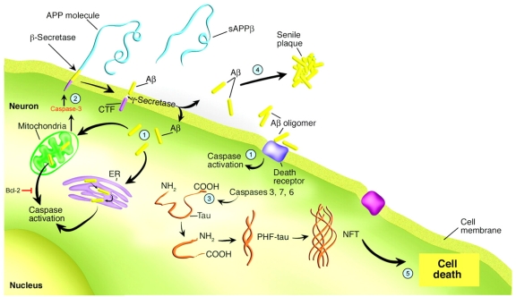
Mounting evidence suggests the involvement of caspases in the disease process associated with Alzheimer's disease (AD). The activation of caspases may be responsible for the neurodegeneration associated with AD and several recent studies have suggested that caspases may also play a role in promoting pathogenic mechanisms underlying this disease. Thus, caspase activation and cleavage of the amyloid precursor protein (APP) and tau may facilitate both the production of beta-amyloid (Abeta) as well as the formation of neurofibrillary tangles (NFTs). Because the activation of caspases in AD may be a proximal event that is not just associated with neurodegeneration, caspases are potential therapeutic targets for the treatment of this disorder. In this review, studies documenting the role of caspases in the AD brain will be discussed. In this context, a discussion of the therapeutic value of targeting caspase inhibition in the treatment of AD will be evaluated including drug targets, delivery and selectivity.

Keywords: Alzheimer’s disease; Apoptosis; Tau; amyloid precursor protein; beta-amyloid; caspase; neurofibrillary tangle.


Figures

Figure 1
Figure 1
The role of caspases in promoting the pathology associated with AD. This figure summarizes the contribution of caspases to the Aβ cascade hypothesis. Step 1: activation of caspases occurs through stimulation of either the receptor-mediated pathway of apoptosis involving cross-linking of death receptors by Aβ (i.e., Fas), through the mitochondrial-mediated Aβ-induced oxidative stress, or finally by ER stress. Step 2: Following the activation of executioner caspases, particularly caspase-3, c-terminal cleavage of APP may facilitate the production of Aβ. This in turn can create a vicious feed-forward cycle of Aβ production and caspase-3 activation. Bcl-2 is an anti-apoptotic protein expressed in mitochondria, which prevents the release of cytochrome c and other pro-apoptotic factors, thus preventing caspase-9 and caspase-3 activation through an interaction with Apaf-1. Step 3: Conversely, caspase-3 cleavage of tau may promote tau aggregation and PHF formation thereby linking Aβ to NFTs. Step 4: Promotion of pathology by the activation of caspases occurs not only through the production of NFTs, but also by leading to Aβ deposition and plaque formation. Step 5: Ultimately, the activation of caspases and cleavage of critical cellular proteins may disrupt axonal and dendritic transport processes leading to cell death and neurodegeneration. Adapted and reproduced by permission from Dickson [27].
Figure 2
Figure 2
Overexpression of Bcl-2 in 3×Tg-AD mice prevents caspase-cleavage and hyperphosphorylation of tau in the amygdala. Panels show representative staining from the amygdala of either 18 month-old 3×Tg-AD mice alone (A and C), or in 3×Tg-AD/Bcl-2 OE mice (B and D). (A and B): representative staining using the tau caspase-cleavage product antibody, an antibody that specifically detects caspase-cleaved tau [25] revealed the presence of caspase-cleaved tau within neurons in 3×Tg-AD mice that was prevented following overexpression of Bcl-2. (C and D): identical to Panels A and B with the exception that an early tangle marker, AT8 was employed. In this case, overexpression of Bcl-2 largely prevented the hyperphosphorylation of tau (D). All scale bars represent 10 μm.

References

    1. Golde TE, Dickson D, Hutton M. Filling the gaps in the abeta cascade hypothesis of Alzheimer's disease. Curr Alzheimer Res. 2006;3:421–430. - PubMed
    1. Christensen DD. Alzheimer's disease: progress in the development of anti-amyloid disease-modifying therapies. CNS Spectr. 2007;12:113–116. 119–123. - PubMed
    1. Su JH, Anderson AJ, Cummings BJ, Cotman CW. Immunohistochemical evidence for DNA fragmentation in neurons in the AD brain. Neuroreport. 1994;5:2529–2533. - PubMed
    1. Anderson AJ, Su JH, Cotman CW. DNA damage and apoptosis in Alzheimer's disease: colocalization with c-Jun immunoreactivity, relationship to brain area, and effect of postmortem delay. J Neurosci. 1996;16:1710–1719. - PMC - PubMed
    1. Gavrieli Y, Sherman Y, Ben-Sasson SA. Identification of programmed cell death in situ via specific labeling of nuclear DNA fragmentation. J Cell Biol. 1992;119:493–501. - PMC - PubMed