. 2009 May-Jun;40(3):14.
doi: 10.1051/vetres:2008052. Epub 2008 Dec 12.

Targeting to porcine sialoadhesin receptor improves antigen presentation to T cells

Affiliations

Targeting to porcine sialoadhesin receptor improves antigen presentation to T cells

Concepción Revilla et al. Vet Res. 2009 May-Jun.

Abstract

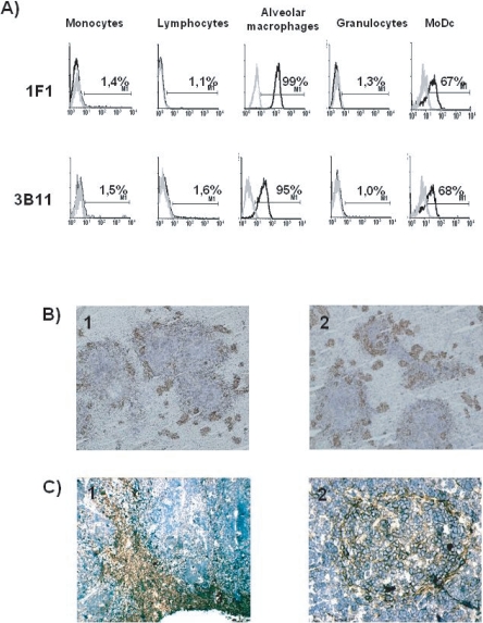
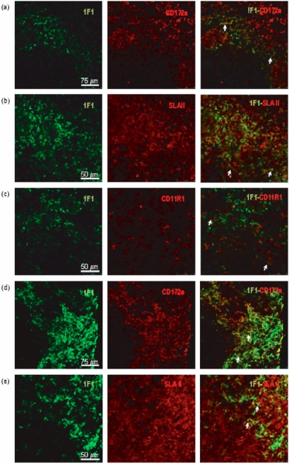
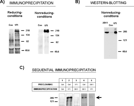
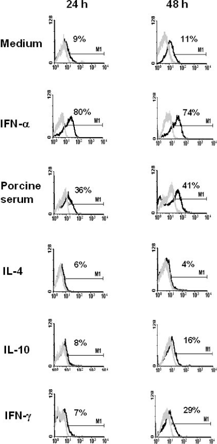
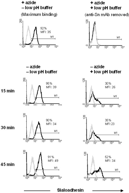
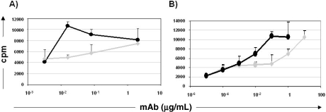
Antibody-mediated targeting of antigen to specific antigen presenting cells (APC) receptors is an attractive strategy to enhance T cell immune responses to weak immunogenic antigens. Here, we describe the characterization of two monoclonal antibodies (mAb) against different epitopes of porcine sialoadhesin (Sn) and evaluate in vitro the potential of targeting this receptor for delivery of antigens to APC for T cell stimulation. The specificity of these mAb was determined by amino acid sequence analysis of peptides derived from the affinity purified antigen. Porcine Sn is expressed by macrophages present in the border between white and red pulp of the spleen and in the subcapsular sinus of lymph nodes, an appropriate location for trapping blood and lymph-borne antigens. It is also expressed by alveolar macrophages and monocyte-derived dendritic cells (MoDC). Blood monocytes are negative for this molecule, but its expression can be induced by treatment with IFN-alpha. MAb bound to Sn is rapidly endocytosed. MAb to sialoadhesin induced in vitro T cell proliferation at concentrations 100-fold lower than the non-targeting control mAb when using T lymphocytes from pigs immunized with mouse immunoglobulins as responder cells and IFN-alpha treated monocytes or MoDC as APC, suggesting a role of sialoadhesin in antigen uptake and/or delivery into the presentation pathway in APC.


Figures

Figure 1.
Figure 1.
(A) MAb 1F1 and 3B11 recognize an antigen expressed in alveolar macrophages and MoDC. Cells were labeled with 1F1 or 3B11 mAb (black histograms) or with irrelevant isotype-matched mAb (grey histograms). Results are representative of five independent experiments. (B) In the spleen, staining with 1F1 (1) and 3B11 (2) mAb appears associated with the walls of the ellipsoidal vessels that surround the islands of white pulp. (C) In lymph nodes, the antigen was detected in the subcapsular sinus and medullary cords (1) and in the periphery of follicles and some follicular cells (2). (A color version of this figure is available at www.vetres.org.)
Figure 2.
Figure 2.
Double staining of spleen (a–c) and mediastinal lymph node (d,e) sections was carried out with 1F1 mAb and mAb to CD172a (a, d), SLAII (b, e) and CD11R1 (c). Arrows point to representative cells co-expressing the indicated markers in the border between white and red pulp of the spleen, and in the subcapsular sinus of lymph nodes.
Figure 3.
Figure 3.
Biochemical characterization of antigen recognized by 1F1 and 3B11 mAbs. (A) Lysates from biotin-labeled alveolar macrophages were immunoprecipitated with 1F1 mAb or an irrelevant isotype-matched control mAb (Con) and analyzed by 7.5% SDS-PAGE under reducing and non-reducing conditions. Numbers in the right indicate position where MW markers ran. (B) Detection by western-blotting of the 1F1 and 3B11 antigens on alveolar macrophage extracts subjected to 7.5% SDS-PAGE under non-reducing conditions. (C) Immunoprecipitation analysis after preclearing. Lysates from biotin-labeled alveolar macrophages were immunoprecipitated with mAb 1F1 and 3B11 after preclearing twice with the mAb indicated in the table. Arrow points the specific band, numbers in the left indicate position where MW markers ran. Results are representative of three independent experiments.
Figure 4.
Figure 4.
MAb 1F1 and 3B11 bind to two different epitopes on the Sn molecule. Black histograms show binding of labeled 3B11 or 1F1 mAb on alveolar macrophages after incubation with the indicated unlabeled antibodies. Grey histograms correspond to the negative control staining. Results are representative of at least two independent experiments using cells from different animals.
Figure 5.
Figure 5.
Regulation of Sn expression on blood monocytes by cytokines or porcine serum. Black histograms show the staining with 1F1 mAb on monocytes subjected to the indicated stimuli after 24 or 48 h. Grey histograms show binding of an irrelevant isotype-matched mAb. Results are representative of five independent experiments.
Figure 6.
Figure 6.
Anti-Sn mAb 1F1 is efficiently internalized in alveolar macrophages upon binding. Black histograms show 1F1- Alexa 488 fluorescence associated to the cells after treatments and 37 °C incubation times indicated in the figure. Cells labeled with 1F1 mAb at 4 °C in the presence of azide to block endocytosis, either untreated (maximum binding) or treated (background binding) with the low pH buffer were used as controls. Grey histograms show binding of an isotype-matched control mAb. Results are representative of three independent experiments.
Figure 7.
Figure 7.
Antigen targeting to Sn enhances T cell immune responses. 3H-thymidine incorporation (cpm) in T cells from pigs immunized with mouse Ig, stimulated with 1F1 mAb (black) or an isotype-matched control mAb (grey) at the indicated concentrations, using as APC: (A) MoDC or (B) Monocytes treated with IFN-a for 24 h. The results are representative of three independent experiments.

References

    1. Berney C., Herren S., Power C.A., Gordon S., Martínez-Pomares L., Kosco-Vilbois M.H.. A member of the dendritic cell family that enters B cell follicles and stimulates primary antibody responses identified by a mannose receptor fusion protein. J. Exp. Med. 1999;190:851–860. - PMC - PubMed
    1. Bimczok D., Post A., Tschernig T., Rothkötter H.-J.. Phenotype and distribution of dendritic cells in the porcine small intestinal and tracheal muchosa and their spatial relationship to epithelial cells. Cell Tissue Res. 2006;325:461–468. - PubMed
    1. Bullido R., Alonso F., Gómez del Moral M., Ezquerra A., Álvarez B., Ortuño E., Domínguez J.. Monoclonal antibody 2F4/11 recognizes the a chain of a porcine β2 integrin involved in adhesion and complement mediated phagocytosis. J. Immunol. Methods. 1996;195:125–134. - PubMed
    1. Bullido R., Gómez del Moral M., Alonso F., Ezquerra A., Zapata A., Sánchez C.. et al. Monoclonal antibodies specific for porcine monocytes/macrophages: macrophage heterogeneity in the pig evidenced by the expression of surface antigens. Tissue Antigens. 1997;49:403–413. - PubMed
    1. Carlin A.F., Lewis A.L., Varki A., Nizet V.. Group B Streptococcal sialic acids interact with Siglecs (immunoglobulin-like lectins) on human leukocytes. J. Bacteriol. 2007;189:1231–1237. - PMC - PubMed

Publication types

LinkOut - more resources