. 2008 Nov;144(5):752-61.
doi: 10.1016/j.surg.2008.07.009. Epub 2008 Sep 2.

Treatment of liver ischemia-reperfusion injury by limiting thrombospondin-1/CD47 signaling

Affiliations

Treatment of liver ischemia-reperfusion injury by limiting thrombospondin-1/CD47 signaling

Jeff S Isenberg et al. Surgery. 2008 Nov.

Abstract

Background: Ischemia-reperfusion (I/R) injury remains a primary complication of transplant surgery, accounting for about 80% of liver transplant failures, and is a major source of morbidity in other pathologic conditions. Activation of endothelium and inflammatory cell recruitment are central to the initiation and promulgation of I/R injury, which can be limited by the bioactive gas nitric oxide (NO). The discovery that thrombsospondin-1 (TSP1), via CD47, limits NO signaling in vascular cells and ischemic injuries in vivo suggested that I/R injury could be another important target of this signaling pathway.

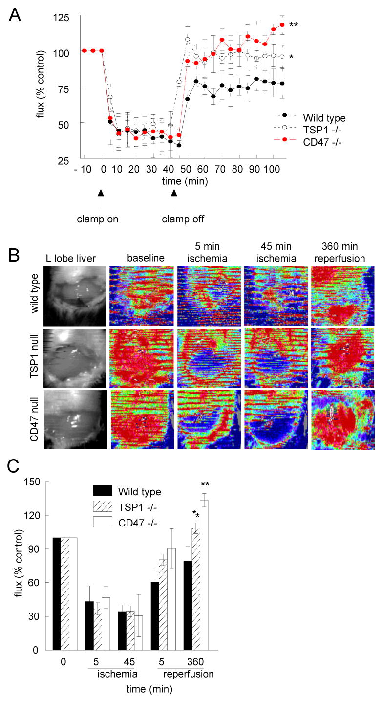
Methods: Wild-type, TSP1-null, and CD47-null mice underwent liver I/R injury. Wild-type animals were pretreated with CD47 or control antibodies before liver I/R injury. Tissue perfusion via laser Doppler imaging, serum enzymes, histology, and immunohistology were assessed.

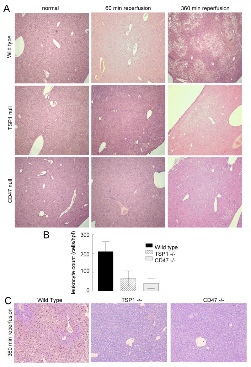
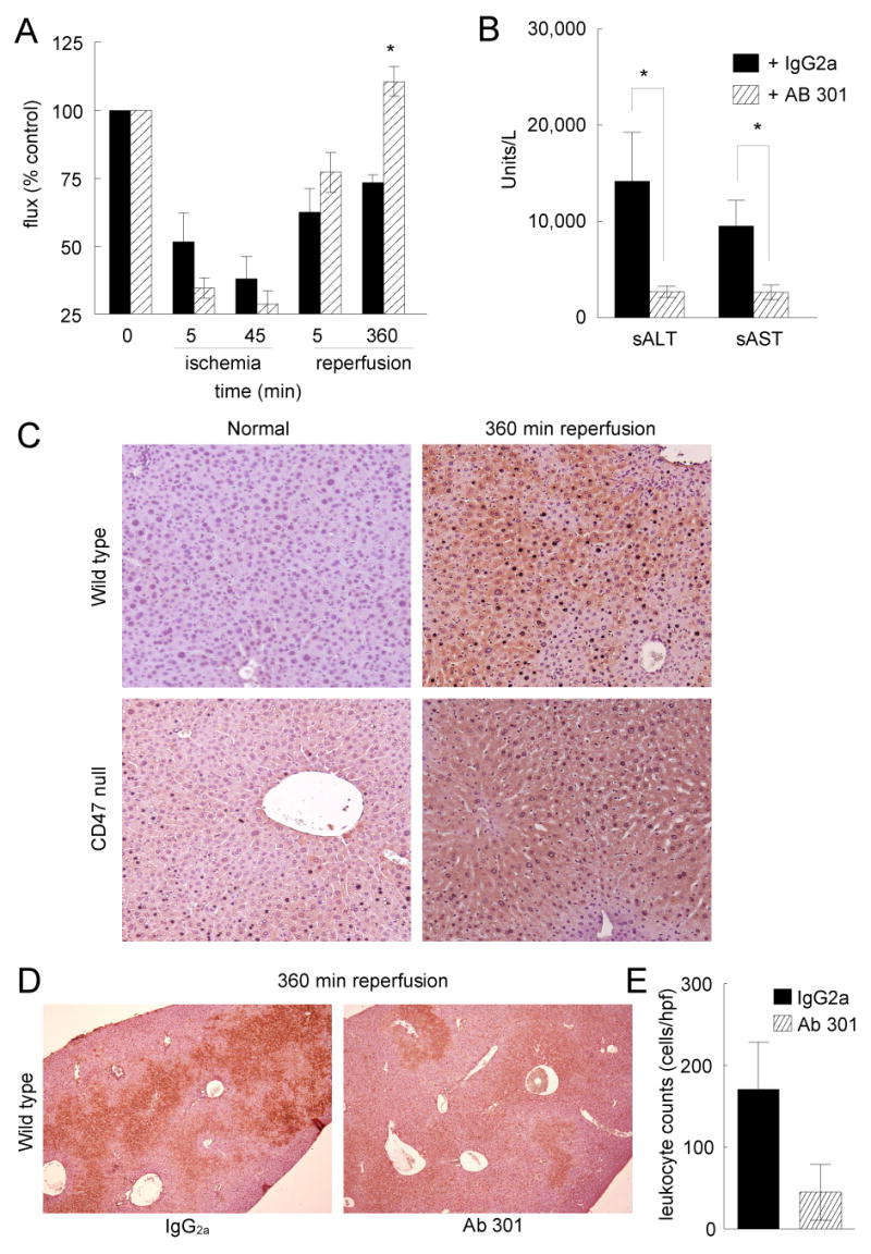
Results: TSP1-null and CD47-null mice subjected to subtotal liver I/R injury showed improved perfusion relative to wild-type mice. Null mice subjected to liver I/R had decreased liver enzyme release and less histologic evidence of injury. Elevated TSP1 expression in liver tissue after I/R injury suggested that preventing its interaction with CD47 could be protective. Thus, pretreatment of wild-type mice using a blocking CD47 antibody improved recovery of tissue perfusion and preserved liver integrity after I/R injury.

Conclusions: Tissue survival and perfusion after liver I/R injury are limited by TSP1 and CD47. Targeting CD47 before I/R injury enhances tissue survival and perfusion in a model of liver I/R injury and suggests therapeutics for enhancing organ survival in transplantation surgery.


Figures

Figure 1
Figure 1. Liver and hind limb reperfusion is limited by TSP1 and CD47
Sex and age matched C57BL/6 wild type, TSP1 null and CD47 null mice underwent 45 min occlusion of the vascular inflow of the left and median hepatic lobes followed by 60 minutes of reperfusion. Laser Doppler analysis of tissue blood flow was performed (A). Results represent the mean ± SD of 12 wild type animals and 6 each of TSP1 null and CD47 null. Data from sham animals is not shown. Sex and age matched C57BL/6 wild type, TSP1 null and CD47 null mice underwent 45 min occlusion of the vascular inflow of the left and median hepatic lobes followed by 360 minutes of reperfusion. Laser Doppler analysis of tissue blood flow was performed. Representative color Doppler images of the liver at baseline, following 5 and 45 minutes of ischemia and 360 minutes of reperfusion are presented (B). Red coloration of laser Doppler images indicates maximum and blue coloration minimum blood flow. Changes in liver perfusion at indicated time points are presented as flux normalized to baseline values (% control, A & C). Results represent the mean ± SD of 12 wild type animals and 6 each of TSP1 null and CD47 null. p < 0.05 TSP1 versus wild type indicated by * and CD47 null versus wild type indicated by **.
Figure 2
Figure 2. Serum enzyme markers are reduced in null animals flowing acute and sub-acute reperfusion
Following liver ischemia and 60 (A) or 360 (B) minutes of reperfusion as described and while maintained at a constant core temperature and under isoflurane anesthesia sex and age matched wild type, TSP1 null and CD47 null mice underwent direct cardiac puncture. Blood was collected in heparin coated syringes, centrifuged, serum separated and immediately processed for serum and liver enzymes. Results represent the mean ± SD of 18 animals, six of each genotype. p < 0.05 CD47 null sALT versus wild type indicated by * and TSP1 null and CD47 null sAST null versus wild type indicated by ** (A) or TSP1 null and CD47 null sALT and sALT versus wild type indicated by * (B).
Figure 3
Figure 3. TSP1 and CD47 null liver sections demonstrate minimal damage following I/R injury
Following 45 min of liver ischemia and either 60 or 360 minutes of reperfusion age and sex matched wild type, TSP1 null and CD47 null mice were euthanized and the left and middle hepatic lobes excised, processed and stained with hematoxylin-eosin (A) (original magnification ×10). Liver sections from sham operated animals were treated similarly. Images are representative liver sections from 18 mice, six of each strain. Inflammatory cell infiltrate was determined in 10 high power fields from wild type, TSP1 null and CD47 null sections (B). Results are from analysis of 72 total sections, 4 sections each from 18 liver lobes, 6 of each genotype. Following 45 min of ischemia and 360 minutes of reperfusion wild type, TSP1 null and CD47 null liver sections were stained for evidence of tissue apoptosis/necrosis (C) (original magnification ×20). Images are representative of sections from 6 mice of each genotype.
Figure 4
Figure 4. Monoclonal antibody targeting of CD47 decreases I/R liver damage
Age and sex matched wild type C57BL/6 mice received either a monoclonal CD47 antibody (clone 301) or an isotype matched control IgG2a antibody 90 minutes pre-operatively. Laser Doppler analysis of liver tissue perfusion was performed pre-operatively, after 45 minutes of ischemia, and following 360 minutes of reperfusion (A). Results represent the mean ± SD of 30 animals, 15 in each treatment group. Blood was collected for analysis of serum liver enzymes (B). Results represent the mean ± SE of 12 animals, six in each treatment group. p < 0.05 IgG2a versus 301 indicated by *. Liver tissue sections were processed for immunohistology. Normal and post I/R injury liver sections (360 minutes reperfusion) from wild type and CD47-null animals (C) (original magnification ×20) or liver sections form wild type animals pretreated with a CD47 antibody (301) or an isotype control IgG2a control antibody prior to I/R injury (360 minutes reperfusion) (D) were stained with a monoclonal TSP1 antibody (clone A6.1) (original magnification ×10). Inflammatory cell infiltrate was determined in 10 high powered fields from wild type, TSP1 null and CD47 null sections (E). Results are from analysis of 48 total sections, 4 sections each from 12 liver lobes, 6 animals in each group.

References

    1. Kupiec-Weglinski JW, Busuttil RW. Ischemia and reperfusion injury in liver transplantation. Transplant Proc. 2005;37(4):1653–6. - PubMed
    1. Kim YI. Ischemia-reperfusion injury of the human liver during hepatic resection. J Hepatobiliary Pancreat Surg. 2003;10(3):195–9. - PubMed
    1. Arii S, Teramoto K, Kawamura T. Current progress in the understanding of and therapeutic strategies for ischemia and reperfusion injury of the liver. J Hepatobiliary Pancreat Surg. 2003;10(3):189–94. - PubMed
    1. Fondevila C, Busuttil RW, Kupiec-Weglinski JW. Hepatic ischemia/reperfusion injury--a fresh look. Exp Mol Pathol. 2003;74(2):86–93. - PubMed
    1. Yin H, Chao L, Chao J. Nitric oxide mediates cardiac protection of tissue kallikrein by reducing inflammation and ventricular remodeling after myocardial ischemia/reperfusion. Life Sci. 2007 - PMC - PubMed

Publication types