. 2008 Dec 10;16(12):1849-59.
doi: 10.1016/j.str.2008.10.010.

Structural basis for inactivation of the human pyruvate dehydrogenase complex by phosphorylation: role of disordered phosphorylation loops

Affiliations

Structural basis for inactivation of the human pyruvate dehydrogenase complex by phosphorylation: role of disordered phosphorylation loops

Masato Kato et al. Structure. .

Abstract

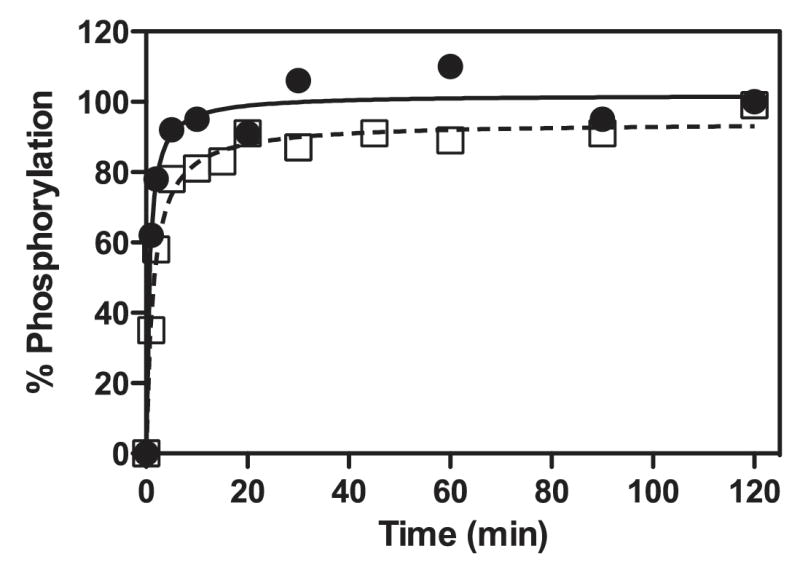
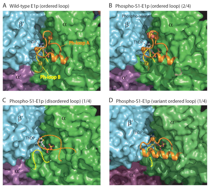
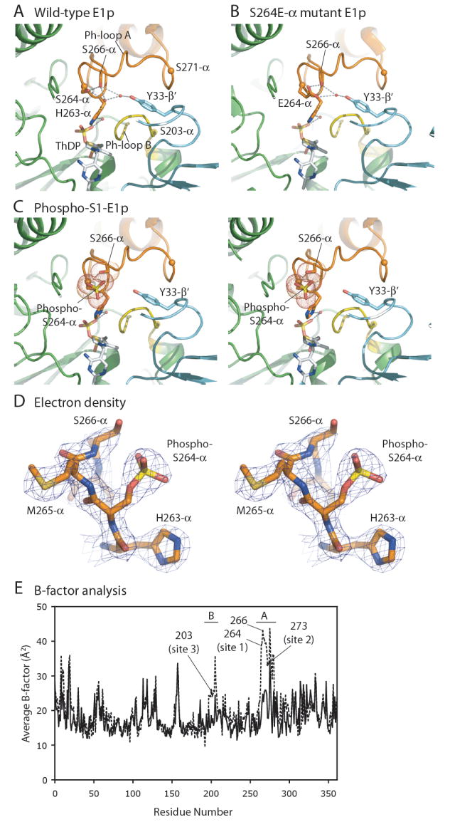
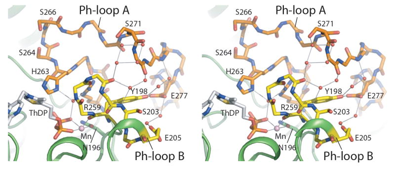
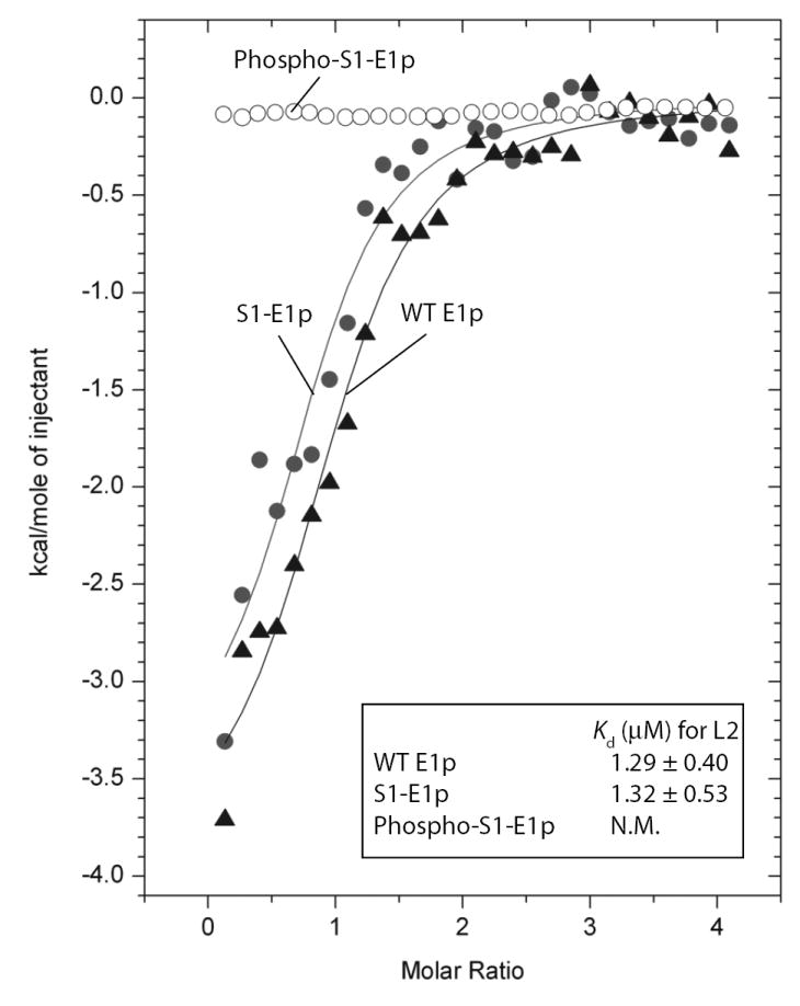
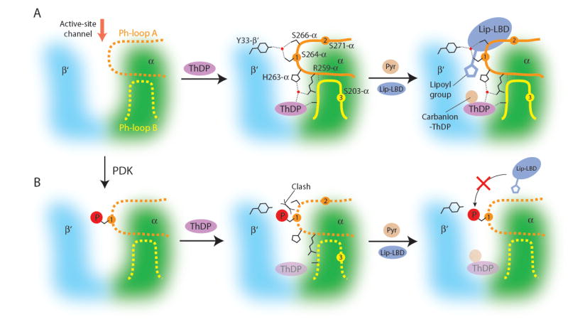
We report the crystal structures of the phosporylated pyruvate dehydrogenase (E1p) component of the human pyruvate dehydrogenase complex (PDC). The complete phosphorylation at Ser264-alpha (site 1) of a variant E1p protein was achieved using robust pyruvate dehydrogenase kinase 4 free of the PDC core. We show that unlike its unmodified counterpart, the presence of a phosphoryl group at Ser264-alpha prevents the cofactor thiamine diphosphate-induced ordering of the two loops carrying the three phosphorylation sites. The disordering of these phosphorylation loops is caused by a previously unrecognized steric clash between the phosphoryl group at site 1 and a nearby Ser266-alpha, which nullifies a hydrogen-bonding network essential for maintaining the loop conformations. The disordered phosphorylation loops impede the binding of lipoyl domains of the PDC core to E1p, negating the reductive acetylation step. This results in the disruption of the substrate channeling in the PDC, leading to the inactivation of this catalytic machine.


Conflict of interest statement

The authors declare no financial conflict of interest related to this work.

Figures

Figure 1
Figure 1. Time Course for Phosphorylation of E1p Variants by PDK4
E1p variants S1-E1p and S2-E1p contain phosphorylatable site 1 and site 2, respectively. The remaining two phosphorylation sites in each E1p variant were mutated to alanine. S1-E1p (●) and S2-E1p (□) were phosphorylated by PDK4 in the absence of the E2p/E3BP cores as described in Experimental Procedures. The amount of [32P] phosphate incorporated per E1p heterotetramer was converted to % phosphorylation. Each variant E1p heterotetramer has two phosphorylatable serine residues, therefore 100% phosphorylation indicates that both serine residues in the heterotetramer are phosphorylated.
Figure 2
Figure 2. Overall Structure of the Wild-type E1p Heterotetramer
A stereo diagram of the crystal structure of the wild-type E1p heterotetramer (α2β2) is shown as surface representation models with each of the four subunits represented by a different color. The two phosphorylation loops (Ph-loops) are shown as ribbon models with Ph-loop A in orange and Ph-loop B in yellow. The three phosphorylation sites in each α subunit are depicted by spheres and labeled according to the site numbers. The red triangle indicates the entrance of the active-site channel where substrates pyruvate and lip-LBD enter. The axis of 2-fold symmetry in the heterotetramer is indicated by a grey arrow.
Figure 3
Figure 3. Structures of the Phosphorylation loops in Wild-type and Phospho-S1-E1p
The structures of Ph-loops in E1p-α subunits are shown as ribbon models against other parts of the protein in surface representation. Ph-loop A (residues from 259-α to 282-α) is in orange, and Ph-loop B (from 198-α to 205-α) in yellow. The three phosphorylation sites are indicated by spheres and labeled according to the site numbers. The bound ThDP is shown as a ball-and-stick model. (A) Fully ordered Ph-loops in wild-type E1p. (B) Wild-type-like ordered Ph-loops in phospho-S1-E1p with bound Mn-ThDP. Two of the four E1p-α subunits in the asymmetric unit (depicted as 2/4 in the figure caption) exhibit this conformation, which is maintained through interactions with a symmetry-related molecule (cf. Figure S3B). (C) Completely disordered Ph-loops in phospho-S1-E1p containing the bound Mn-ThDP. One of the four E1p-α subunit in the asymmetric unit (1/4) has this conformation. No symmetry-related molecule is present near the Ph-loops (cf. Figure S3A). (D) Variant ordered conformation of Ph-loop A and partially ordered Ph-loop B in the remaining E1p-α subunits (1/4). This conformation is also maintained by interactions with a symmetry-related molecule (cf. Figure S3C).
Figure 4
Figure 4. Hydrogen-Bond Networks Involving Phosphorylation Site 1
(A) The H-bond network connecting phosphorylation site 1 and Tyr33-β’ of the E1p-β’ subunit in the wild-type E1p structure. Ph-loop A is in orange, and Ph-loop B in yellow. The positions of the three phosphorylation sites are shown as spheres at the corresponding Cα atom positions. Water molecules are depicted as small red dots. H-bonds are indicated by grey dashed lines. (B) A similar H-bond network in the Ser264E-α mutant E1p structure (PDB code: 2OZL) (Seifert et al., 2007). (C) A stereo diagram showing absence of the H-bond network in phospho-S1-E1p containing bound Mn-ThDP. The phosphoryl group on Ser264-α (site 1) clashes with the side chain of the neighboring Ser266-α. Van der Waals radii of the phosphoryl group and side chains of both serine residues are shown as spheres of red dots. (D) A stereo figure of the 2Fo-Fc electron density map (contoured at 1σ) at phosphorylation site 1 with a stick representation of the refined model. (E) Average B-factor plots of the wild-type and phospho-S1-E1p structures. Average B-factors for individual residues in one of the four non-phosphorylated wild-type E1p-α subunits (solid line) and one of the two phospho-S1-E1p-α subunits with the wild-type-like “ordered” Ph-loops (dashed line) are plotted against the residue number. The residue ranges for Ph-loops A and B are indicated on top of the graph. Each of the three phosphorylation sites and residue Ser266-α are labeled.
Figure 5
Figure 5. Extensive Interactions Between the Two Ph-loops in the Wild-Type E1p Structure
The stereo diagram illustrates extensive interactions between Ph-loop A (orange) and Ph-loop B (yellow) in the non-phosphorylated wild-type E1p structure. The Ph-loops are shown as stick models. Side chains of some residues are omitted for clarity. The three phosphorylation sites are S264-α (site 1), S271-α (site 2), and S203-α (site 3). Water molecules are shown as small red spheres and the manganese atom as a pink sphere. H-bonds are indicated by grey lines.
Figure 6
Figure 6. Binding of the Lipoylated-L2 Domain to E1p Determined by Isothermal Titration Calorimetry
Wild-type E1p, S1-E1p or phospho-S1-E1p protein (35 μM, heterodimer) was titrated with 0.65 mM lip-L2 (residues 128-265 in E2p) as described in the Experimental Procedures. The binding isotherms were fit using ORIGIN v. 7.0 software. For wild-type E1p and S1-E1p, the dissociation constants Kd are averages of triplicates; for phospho-S1-E1p, there are no detectable heats.
Figure 7
Figure 7. Mechanism of PDC Inactivation by Site 1 Phosphorylation in E1p
Reaction scheme for non-phosphorylated E1p. The E1p active site is formed by portions of the α subunit (green) and the β’ subunit (cyan). The red arrow depicts the entrance of the active-site channel for substrates pyruvate and lip-LBD. In apo E1p, Ph-loop A (orange) and Ph-loop B (yellow) are disordered (shown as dashed lines in the left panel). Binding of the cofactor ThDP (magenta) at the bottom of the active-site channel induces ordering of both Ph-loop A and Ph-loop B (middle panel). Phosphorylation sites are indicated by orange and yellow circles and numbered. Water molecules are represented by red dots. H-bonds are shown as dashed lines. These ordered Ph-loops mediate E1p-catalyzed decarboxylation of pyruvate and reductive acetylation of lip-LBD (right panel). (B) Inactivation mechanism for phosphorylated E1p. When Ph-loop A is disordered in apo E1p, Ser264-α (site 1) is phosphorylatable by PDK (from panel A left to panel B left). A steric clash between the phosphoryl group and adjacent Ser266-α prevents the ordering of Ph-loops (middle panel). In the absence of the ordered Ph-loops, ThDP binding to the E1p active site is significantly attenuated (shown as a transparent oval). Nonetheless, under saturating ThDP conditions, the E1p-catalyzed decarboxylation of pyruvate proceeds (right panel). However, the reductive acetylation of lip-LBD is abolished due to the loss of determinants on the disordered Ph-loops, which are essential for lip-LBD binding to the E1p active site. The abrogation of E1p-catalyzed reductive acetylation interrupts substrate channeling in the PDC, resulting in the inactivation of this catalytic machine.

References

    1. Ali MS, Shenoy BC, Eswaran D, Andersson LA, Roche TE, Patel MS. Identification of the tryptophan residue in the thiamin pyrophosphate binding site of mammalian pyruvate dehydrogenase. J Biol Chem. 1995;270:4570–4574. - PubMed
    1. Bao H, Kasten SA, Yan X, Hiromasa Y, Roche TE. Pyruvate Dehydrogenase Kinase Isoform 2 Activity Stimulated by Speeding Up the Rate of Dissociation of ADP. Biochemistry. 2004;43:13442–13451. - PubMed
    1. Bonnet S, Archer SL, Allalunis-Turner J, Haromy A, Beaulieu C, Thompson R, Lee CT, Lopaschuk GD, Puttagunta L, Bonnet S, et al. A mitochondria-K+ channel axis is suppressed in cancer and its normalization promotes apoptosis and inhibits cancer growth. Cancer cell. 2007;11:37–51. - PubMed
    1. Cairns RA, Papandreou I, Sutphin PD, Denko NC. Metabolic targeting of hypoxia and HIF1 in solid tumors can enhance cytotoxic chemotherapy. PNAS. 2007;104:9445–9450. - PMC - PubMed
    1. CCP4. The CCP4 suite: programs for protein crystallography. Acta Crystallogr D Biol Crystallogr. 1994;50:760–763. - PubMed

Publication types

MeSH terms