. 2008 Dec;15(6):901-12.
doi: 10.1016/j.devcel.2008.09.018.

The conserved protein SZY-20 opposes the Plk4-related kinase ZYG-1 to limit centrosome size

Affiliations

The conserved protein SZY-20 opposes the Plk4-related kinase ZYG-1 to limit centrosome size

Mi Hye Song et al. Dev Cell. 2008 Dec.

Abstract

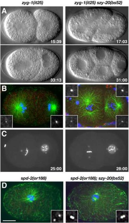
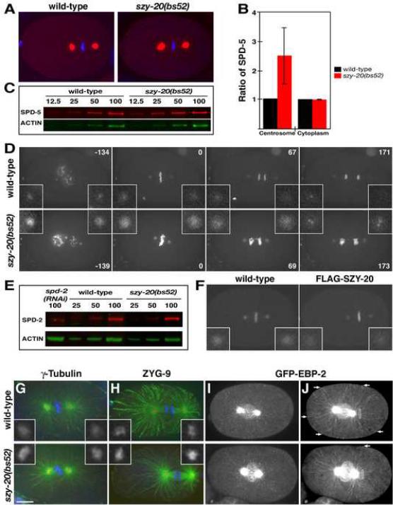
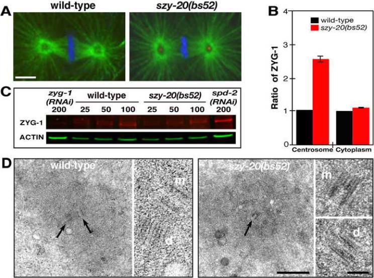
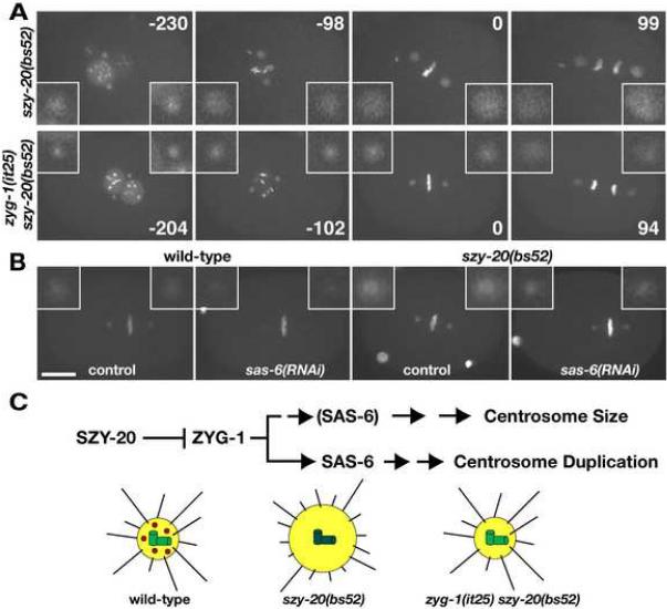
Microtubules are organized by the centrosome, a dynamic organelle that exhibits changes in both size and number during the cell cycle. Here we show that SZY-20, a putative RNA-binding protein, plays a critical role in limiting centrosome size in C. elegans. SZY-20 localizes in part to centrosomes and in its absence centrosomes possess increased levels of centriolar and pericentriolar components including gamma-tubulin and the centriole duplication factors ZYG-1 and SPD-2. These enlarged centrosomes possess normal centrioles, nucleate more microtubules, and fail to properly direct a number of microtubule-dependent processes. Depletion of ZYG-1 restores normal centrosome size and function to szy-20 mutants, whereas loss of szy-20 suppresses the centrosome duplication defects in both zyg-1 and spd-2 mutants. Our results describe a pathway that determines centrosome size and implicate centriole duplication factors in this process.


Figures

Figure 1
Figure 1
Loss of szy-20 activity suppresses centrosome duplication defects in zyg-1 and spd-2 mutants. (A–C) Centrosome duplication and bipolar spindle formation are restored in zyg-1(it25) szy-20(bs52) embryos. (A) Selected images from 4D-DIC recordings. Only the double mutant embryo progresses to the four-cell stage. (B) Two-cell stage embryos stained for microtubules (green), centrioles (SAS-4, red) and DNA (blue). (C) Selected images from recordings of embryos expressing GFP-SPD-2 and GFP-histone. Time (minutes) is relative to first metaphase. (D) One-cell embryos stained for microtubules (green), SAS-4 (red) and DNA (blue). The spd-2(or188) embryo has two centrioles and the spd-2(or188) szy-20(bs52) embryo four. Each image is a Z-projection. Insets are magnified 3-fold. Bar, 10 μm.
Figure 2
Figure 2
SZY-20 is required for cell division. szy-20(bs52) embryos stained for microtubules (green), centrosomes (red) and DNA (blue). (A) A one-cell embryo with four centrosomes stained for ZYG-1. (B) A two-cell embryo with detached centrosomes (arrows) stained for SPD-2. (C) A zygote with two sperm pronuclei and centrosomes (boxed) stained for SAS-4. Each image is Z-projection. Insets are magnified 3-fold. Bar, 10 μm.
Figure 3
Figure 3
szy-20 encodes a novel conserved protein. (A) Schematic of wild-type and mutant forms of SZY-20. Due to a frame shift, the product of the szy-20(tm1997) allele is expected to contain a novel 36-amino acid extension (blue box). (B) Immunoblot of wild-type and szy-20(bs52) embryonic extracts probed for SZY-20 (red), and α-tubulin (green) as a loading control. (C–G) SZY-20 localizes to the centrosome. Embryos stained for microtubules (green), SZY-20 (αS20N, red) and DNA (blue): Wild-type embryos at (C) meiosis, (D) first metaphase, (E) first anaphase, (F) second interphase, and (H) a szy-20(RNAi) embryo at metaphase. (G) A metaphase embryo co-stained for SZY-20 (red) and GFP-SAS-4 (green). Boxes highlight centrosomes. Insets are magnified 3-fold. Bar, 10 μm.
Figure 4
Figure 4
SZY-20 limits the size and the microtubule-nucleating capacity of the centrosome. (A) Embryos at first metaphase stained for SPD-5 (red) and DNA (blue). (B) Fluorescence intensity measurements of centrosomal and cytoplasmic SPD-5. Vertical bars indicate the standard deviation. (C) Quantitative immunoblot of SPD-5. Relative loading volumes are indicated above lanes. Actin served as a loading control (D) Still images from recordings of embryos expressing GFP-SPD-2 and GFP-histone. Note that in the szy-20(bs52) embryo, more chromosomes are present due to polar body extrusion failure and that the metaphase spindle is shorter. Time (in seconds) is relative to metaphase. (E) Quantitative immunoblot of SPD-2. (F) Overexpression of wild-type FLAG-SZY-20 reduces GFP-SPD-2 fluorescence at centrosomes. (G, H) Embryos stained for microtubules (green), centrosomes (red) and DNA (blue). The levels of (G) γ-tubulin and (H) ZYG-9 are increased at szy-20(bs52) centrosomes compared to the wild type. (I, J) Time-lapse recordings of embryos expressing GFP-EBP-2. (I) A Z-projection of a single time point illustrates the presence of more GFP-EBP-2 at szy-20(bs52) centrosomes. (J) Time projections of a 10-second interval show the tracks of growing microtubules. During this period, more microtubules grow to reach the cortex (arrows) in the wild type than in the mutant. Boxes (6 μm2) indicate area used to measure GFP-EBP-2 fluorescence. Each image is a Z-projection. Insets are magnified 3-fold. Bar, 10 μm.
Figure 5
Figure 5
Loss of SZY-20 activity enhances ZYG-1 localization at centrosomes. (A) a Z-projections of embryos at first metaphase stained for microtubules (green), ZYG-1 (red) and DNA (blue). ZYG-1 is detectable only at szy-20(bs52) centrosomes. Bar, 5 μm. (B) Fluorescence intensity measurements of centrosomal and cytoplasmic ZYG-1. Vertical bars indicate the standard deviation. (C) Quantitative immunoblot of ZYG-1. Relative loading volumes are indicated above lanes. Actin served as a loading control. (D) TEM of centrosomes at anaphase. In the large panels, a wild-type centriole pair (arrows) and the mother centriole (arrow) of a szy-20(bs52) centriole pair are shown. In the small panels, all mother (m) and daughter (d) centrioles are shown at higher magnification. Bars, 500 nm for lower and 100 nm for higher magnification images.
Figure 6
Figure 6
Centriole duplication factors regulate centrosome size. (A, B) Images from recordings of embryos expressing GFP-SPD-2 and GFP-histone. (A) Reducing zyg-1 activity restores normal centrosome size and function to szy-20(bs52) mutants. Note that the delay in pronuclear rotation observed in szy-20(bs52) is rescued by the zyg-1(it25) mutation. Time (seconds) is relative to first metaphase. (B) sas-6(RNAi) reduces centrosome size in both wild-type and szy-20(bs52) embryos. Shown are Z-projections at each time point. Insets are magnified 3-fold. Bar, 10 μm. (C) A molecular pathway for controlling centrosome size (Top). SZY-20 acts upstream and controls the centrosomal level of ZYG-1, which regulates both centrosome duplication and size. The centriole duplication factor SAS-6 also influences centrosome size. It is shown in parentheses in the `Centrosome Size' Pathway to indicate that its role here has not yet been firmly established. A depiction of the effects of SZY-20 on the centrosome (Bottom). In the wild type, SZY-20 (red) restricts the amount of ZYG-1 (green) at centrioles, thereby establishing the proper level of PCM (yellow) and microtubules (black lines). In szy-20 mutants, ZYG-1 levels at centrioles increase, leading to enhanced recruitment of PCM and the nucleation of more microtubules (on average these are shorter than those nucleated by wild-type centrosomes). Reducing ZYG-1 activity in szy-20 mutants restores normal levels of PCM and microtubule-nucleation.

References

    1. Alliegro MC, Alliegro MA, Palazzo RE. Centrosome-associated RNA in surf clam oocytes. Proc Natl Acad Sci U S A. 2006;103:9034–9038. - PMC - PubMed
    1. Altschul SF, Madden TL, Schaffer AA, Zhang J, Zhang Z, Miller W, Lipman DJ. Gapped BLAST and PSI-BLAST: a new generation of protein database search programs. Nucleic Acids Res. 1997;25:3389–3402. - PMC - PubMed
    1. Azimzadeh J, Bornens M. Structure and duplication of the centrosome. J Cell Sci. 2007;120:2139–2142. - PubMed
    1. Bettencourt-Dias M, Rodrigues-Martins A, Carpenter L, Riparbelli M, Lehmann L, Gatt MK, Carmo N, Balloux F, Callaini G, Glover DM. SAK/PLK4 is required for centriole duplication and flagella development. Curr Biol. 2005;15:2199–2207. - PubMed
    1. Blower MD, Feric E, Weis K, Heald R. Genome-wide analysis demonstrates conserved localization of messenger RNAs to mitotic microtubules. J Cell Biol. 2007;179:1365–1373. - PMC - PubMed

Publication types

MeSH terms

LinkOut - more resources