Review
. 2008 Dec 10;60(5):748-66.
doi: 10.1016/j.neuron.2008.10.010.

Mitochondria in neuroplasticity and neurological disorders

Affiliations
Review

Mitochondria in neuroplasticity and neurological disorders

Mark P Mattson et al. Neuron. .

Abstract

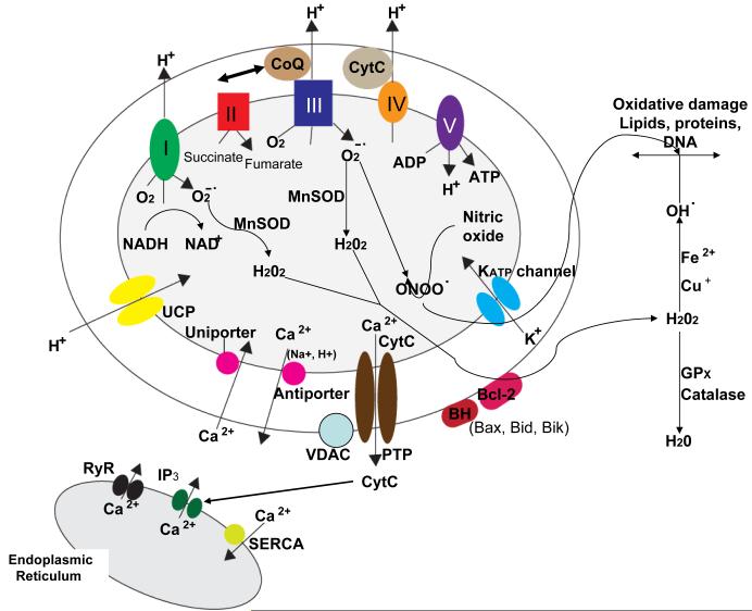
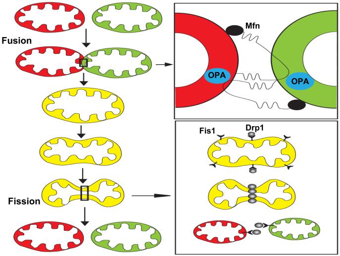
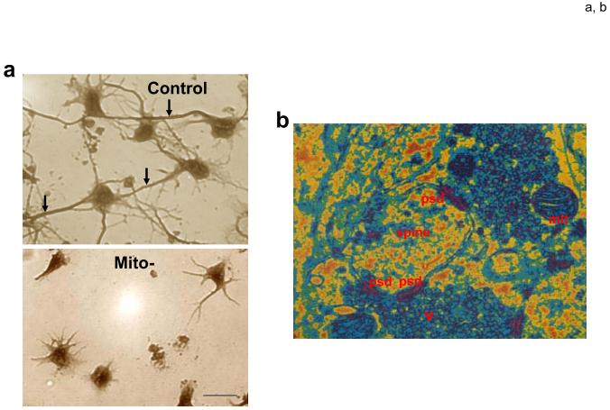
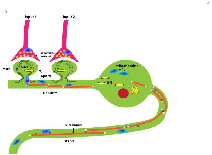
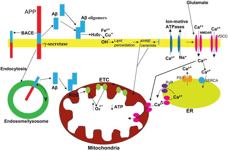
Mitochondrial electron transport generates the ATP that is essential for the excitability and survival of neurons, and the protein phosphorylation reactions that mediate synaptic signaling and related long-term changes in neuronal structure and function. Mitochondria are highly dynamic organelles that divide, fuse, and move purposefully within axons and dendrites. Major functions of mitochondria in neurons include the regulation of Ca(2+) and redox signaling, developmental and synaptic plasticity, and the arbitration of cell survival and death. The importance of mitochondria in neurons is evident in the neurological phenotypes in rare diseases caused by mutations in mitochondrial genes. Mitochondria-mediated oxidative stress, perturbed Ca(2+) homeostasis, and apoptosis may also contribute to the pathogenesis of prominent neurological diseases including Alzheimer's, Parkinson's, and Huntington's diseases; stroke; amyotrophic lateral sclerosis; and psychiatric disorders. Advances in understanding the molecular and cell biology of mitochondria are leading to novel approaches for the prevention and treatment of neurological disorders.


Figures

Figure 1
Figure 1
Proteins involved in mitochondrial bioenergetics, oxygen radical metabolism and Ca2+ regulation. The electron transport chain consists of four protein complexes (I-IV) and the ATP synthase (complex V) located in the mitochondrial inner membrane. The activity of complex I converts NADH to NAD+ and the activity of complex II converts succinate to fumarate. Complexes I, III and IV transport protons (H+) across the membrane and complexes I and III generate superoxide anion radical (O2-.) during the electron transfer process. The enzymatic activity of mitochondrial manganese superoxide dismutase (MnSOD) converts O2-. to hydrogen peroxide (H2O2) which may then diffuse to the cytoplasmic compartments where glutathione peroxidase and catalase convert H2O2 to H2O. However, H2O2 can interact with Fe2+ or Cu+ to generate hydroxyl radical (OH.), a highly reactive free radical that can induce lipid peroxidation and oxidative damage to proteins and DNA. Mitochondrial uncoupling proteins (UCP) function as H+ leak channels which decrease mitochondrial membrane potential results in decreased generation of O2-. and ATP. Several mitochondrial proteins are involved in regulating movement of Ca2+ into and out of the mitochondria including the Ca2+ uniporter which moves Ca2+ into the mitochondrial matrix and the Ca2+ antiporter which extrudes Ca2+ into the cytosol. In addition, movement of K+ through ATP-sensitive potassium channels (KATP) in the inner membrane can result in decreased mitochondrial Ca2+ uptake. An important transmembrane protein complex which includes the voltage-dependent anion channel (VDAC) forms large permeability transition pores (PTP). The PTP open during the process of apoptosis resulting in the release of cytochrome c into the cytoplasm. Several cytoplasmic proteins may also interact with mitochondrial membranes resulting in a change its permeability including Bcl-2 and BH-only proteins such as Bax, Bid and Bik. Finally, there are interactions between mitochondria and the endoplasmic reticulum (ER) such that Ca2+ released through ER IP3 receptors and ryanodine receptors (RyR) is rapidly transferred into mitochondria. On the other hand, cytochrome c released from mitochondria can trigger the release of Ca2+ from the ER.
Figure 2
Figure 2
Mitochondrial fusion and fission mechanisms. Mitochondria can fuse with each other and exchange their membrane, intermembrane and matrix components (upper left). Fusion of the outer mitochondrial membranes is mediated by homophilic and heterophilic interactions of cytosolic domains of mitofusins (Mfn1 and Mfn2) which are proteins located in the outer mitochondrial membrane (upper right). Fusion of the inner mitochondrial membranes is believed to be mediated by OPA1 which is located in the intermembrane space. Mitochondrial fission (lower left) involves recruitment of dynamin-related protein 1 (Drp1) to discrete foci within the mitochondria and also requires Fis1, a protein located in the outer mitochondrial membrane (lower right).
Figure 3
Figure 3
Mitochondrial trafficking mechanisms within the axon. Microtubules serve as tracks along which mitochondria move either towards the presynaptic terminal (anterograde transport) or towards the cell body (retrograde transport). ATP-dependent motor proteins that move mitochondria along microtubules include kinesins (anterograde) and dynein (retrograde). Mitochondria associate with the motor proteins through specific adaptor proteins. Adaptor proteins (AP) for kinesins include Milton, syntabulin and a Rho GTPase called Miro. Dynactin is an adaptor protein for dynein. Within the axonal growth cone and presynaptic terminal, mitochondria may be anchored and moved along actin filaments by a myosin-mediated mechanism.
Figure 4
Figure 4
Roles of mitochondria in developmental and synaptic plasticity. a. Mitochondria play a pivotal role in axogenesis. At the time of plating in cell culture, embryonic rat hippocampal neurons were not (control) or were (Mito-) treated with ethidium bromide which damages mitochondrial DNA thereby rendering the ETC dysfunctional. Images show neurons approximately 4 days later; the neurons in the control culture elaborated long axons (arrows) and shorter dendrites, whereas the Mito- neurons formed only shorter process and no axon. b. Electron microscopic image of synapses in the adult rodent brain showing a dendritic spine with three postsynaptic densities (psd), two presynaptic terminals with numerous synaptic vesicles (v) and a mitochondrion (mit) in one presynaptic terminal. c. Involvement of mitochondrial motility in synaptic plasticity. This example shows a neuron receiving synaptic inputs onto two dendritic spines. Activation of Input 1 results in Ca2+ influx into the dendritic spine which induces the local engagement of cytoskeleton-mitochondria interactions resulting in the translocation of a mitochondrion to the base of that spine. In contrast, mitochondria are not recruited to an adjacent inactive synapse (Input 2). Mitochondrial transport along axons and dendrites may also be influenced by action potentials. By moving to regions of active synapses mitochondria may contribute to plasticity by increasing the local supply of ATP and by buffering and releasing Ca2+. ER, endoplasmic reticulum.
Figure 4
Figure 4
Roles of mitochondria in developmental and synaptic plasticity. a. Mitochondria play a pivotal role in axogenesis. At the time of plating in cell culture, embryonic rat hippocampal neurons were not (control) or were (Mito-) treated with ethidium bromide which damages mitochondrial DNA thereby rendering the ETC dysfunctional. Images show neurons approximately 4 days later; the neurons in the control culture elaborated long axons (arrows) and shorter dendrites, whereas the Mito- neurons formed only shorter process and no axon. b. Electron microscopic image of synapses in the adult rodent brain showing a dendritic spine with three postsynaptic densities (psd), two presynaptic terminals with numerous synaptic vesicles (v) and a mitochondrion (mit) in one presynaptic terminal. c. Involvement of mitochondrial motility in synaptic plasticity. This example shows a neuron receiving synaptic inputs onto two dendritic spines. Activation of Input 1 results in Ca2+ influx into the dendritic spine which induces the local engagement of cytoskeleton-mitochondria interactions resulting in the translocation of a mitochondrion to the base of that spine. In contrast, mitochondria are not recruited to an adjacent inactive synapse (Input 2). Mitochondrial transport along axons and dendrites may also be influenced by action potentials. By moving to regions of active synapses mitochondria may contribute to plasticity by increasing the local supply of ATP and by buffering and releasing Ca2+. ER, endoplasmic reticulum.
Figure 5
Figure 5
Involvement of amyloidogenic APP processing, oxidative stress and perturbed cellular Ca2+ homeostasis in mitochondrial dysfunction in Alzheimer’s disease. Amyloidogenic processing of the β-amyloid precursor protein (APP) involves sequential cleavages by β-secretase (BACE) which cleaves APP at the cell surface and γ-secretase which cleaves within the membrane-spanning domain of APP resulting in the liberation of the amyloid β-peptide (Aβ). Aβ monomers interact to form oligomers and during this process Aβ may interact with Fe2+ or Cu+ to generate H2O2 and OH. resulting in membrane lipid peroxidation and the generation of 4-hydroxynonenal (HNE) and ceramide. By impairing the function of plasma membrane ion-motive ATPases (Na+ and Ca2+ pumps) and glucose and glutamate transporters (not shown) HNE promotes excessive Ca2+ influx through N-methyl-D-aspartate (NMDA) receptors and voltage-dependent Ca2+ channels (VDCC). Ab may also form Ca2+-conducting pores in the plasma membrane. Excessive Ca2+ influx and release from endoplasmic reticulum (ER) stores may then result in excessive Ca2+ uptake into mitochondria and impairment of their function. Alternatively, HNE and ceramide may diffuse to mitochondria and directly damage mitochondrial membranes. Aβ may also be generated intracellularly in an endosomal/lysosomal compartment; intracellular Aβ may interact with and damage mitochondrial membranes. In these ways, Aβ may impair mitochondrial ATP production and Ca2+ regulation, with adverse consequences for neuronal plasticity and survival.
Figure 6
Figure 6
The degeneration of dopaminergic neurons in Parkinson’s disease involves impaired ETC function, proteasomal overload and excitotoxicity. Mitochondrial complex I activity is reduced in vulnerable neurons in PD, likely as the result of a combination of normal aging, exposures to environmental toxins and genetic factors. The resulting ATP depletion and increased levels of ROS render neurons vulnerable to excitotoxic Ca2+ overload. Mutations in genes that cause inherited PD (α-synuclein, Parkin, DJ-1, PINK1, UCHL1 and LRRK2) may adversely affect mitochondrial function either indirectly or directly. Mutations of α-synuclein (or increased amounts of wild-type α-synuclein caused by increased expression or decreased proteasomal degradation) results in the formation of α-synuclein oligomers which may exacerbate ROS-mediated damage to mitochondrial membranes and proteins. UCHL2 mutations may contribute to proteasomal overload in PD. Parkin is a ubiquitin E3 ligase that plays important roles in removing damaged proteins from neurons; this E3 ligase activity is reduced in PD resulting in excessive accumulation of damaged/neurotoxic proteins. Parkin may affect one or more proteins of the PTP, thereby preventing cytochrome c release and apoptosis. DJ-1 is a mitochondrial protein that reduces ROS and blocks PTP formation. PINK1 is important for the maintenance of membrane potential and suppression of oxidative stress. Thus, mutations in DJ-1 and PINK1 promote damage to mitochondria. E1, ubiquitin E1 ligase; E2, ubiquitin E2 ligase; LRRK2, leucine-rich repeat kinase 2; UCHL1, ubiquitin C-terminal hydrolase L1.

Similar articles

Cited by

References

    1. Abeliovich A, Schmitz Y, Fariñas I, Choi-Lundberg D, Ho WH, Castillo PE, Shinsky N, Verdugo JM, Armanini M, Ryan A, et al. Mice lacking alpha-synuclein display functional deficits in the nigrostriatal dopamine system. Neuron. 2000;25:239–252. - PubMed
    1. Andres-Mateos E, Perier C, Zhang L, Blanchard-Fillion B, Greco TM, Thomas B, Ko HS, Sasaki M, Ischiropoulos H, Przedborski S, et al. DJ-1 gene deletion reveals that DJ-1 is an atypical peroxiredoxin-like peroxidase. Proc. Natl. Acad. Sci. U. S. A. 2007;104:14807–14812. - PMC - PubMed
    1. Arthur PG, Matich GP, Pang WW, Yu DY, Bogoyevitch MA. Necrotic death of neurons following an excitotoxic insult is prevented by a peptide inhibitor of c-jun N-terminal kinase. J. Neurochem. 2007;102:65–76. - PubMed
    1. Arumugam TV, Gleichmann M, Tang SC, Mattson MP. Hormesis/preconditioning mechanisms, the nervous system and aging. Ageing Res. Rev. 2006;5:165–178. - PubMed
    1. Atsumi T. The ultrastructure of intramuscular nerves in amyotrophic lateral sclerosis. Acta Neuropathol. 1981;55:193–198. - PubMed

Publication types

MeSH terms

Substances