Review
. 2009 Feb 20;384(2):324-34.
doi: 10.1016/j.virol.2008.11.017. Epub 2008 Dec 10.

Papillomavirus E6 proteins

Affiliations
Review

Papillomavirus E6 proteins

Heather L Howie et al. Virology. .

Abstract

The papillomaviruses are small DNA viruses that encode approximately eight genes, and require the host cell DNA replication machinery for their viral DNA replication. Thus papillomaviruses have evolved strategies to induce host cell DNA synthesis balanced with strategies to protect the cell from unscheduled replication. While the papillomavirus E1 and E2 genes are directly involved in viral replication by binding to and unwinding the origin of replication, the E6 and E7 proteins have auxillary functions that promote proliferation. As a consequence of disrupting the normal checkpoints that regulate cell cycle entry and progression, the E6 and E7 proteins play a key role in the oncogenic properties of human papillomaviruses with a high risk of causing anogenital cancers (HR HPVs). As a consequence, E6 and E7 of HR HPVs are invariably expressed in cervical cancers. This article will focus on the E6 protein and its numerous activities including inactivating p53, blocking apoptosis, activating telomerase, disrupting cell adhesion, polarity and epithelial differentiation, altering transcription and reducing immune recognition.
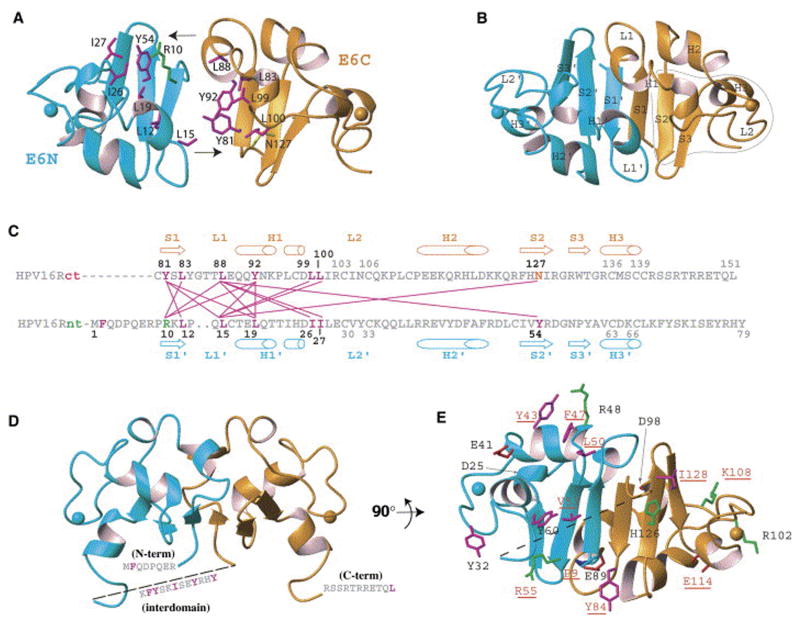

Figures

Figure 1
Figure 1
The Pseudodimeric Model of E6 (taken from Nominé et al, 2006). (A) E6N model and E6C structure positioned symmetrically with exposed hydrophobic patches facing each other. (B) Proposed pseudodimeric arrangement of E6N and E6C. The gray closed line indicates the DNA binding region of E6C, mapped in the present work. (C) Contact map of hydrophobic residues involved in the interface of the pseudodimeric model. (D) Position of the three flanking regions not predicted by the model.(E) A view of the core model highlighting surface residues conserved in all E6 proteins. Previously mutated residues are indicated with underlined red labels.
Figure 2
Figure 2
Binding Partners of E6. E6 alters numerous cellular pathways through the binding of other proteins.
Figure 3
Figure 3
Extrinsic and Intrinsic Apoptotic Pathways. The extrinsic pathway is activated through trimerization of death receptors, followed by recruitment of adapter molecules and pro-caspase 8 to form the DISC. Subsequent cleavage of caspase 8 leads to cleavage of downstream executioner caspases, caspase 3 and caspase 7, and subsequent cell death. E6 can block death receptor association with adapter molecules (1) and can induce the degradation of certain adapter molecules as well as caspase 8 (2). Activation of the intrinsic pathway by “intrinsic” cell stresses leads to the formation of pores in the mitochondrial membrane, and subsequent release of mitochondrial inner membrane proteins into the cytosol. These proteins, along with pro-caspase 9 form the apoptosome, which cleaves caspase 9. Activated caspase 9 then goes on to cleave the downstream executioner caspases 3 and 7, leading to cell death. E6 has been shown to degrade Bak, and thus block the release of the mitochondrial inner membrane proteins into the cytosol (3). E6 is also able to inhibit IAPs and thus block the apoptosome and cleavage of the exectutioner caspases (4).
Figure 4
Figure 4
Model of the hTERT promoter. The catalytic subunit of telomerase, hTERT, is regulated at its core promoter by cis elements that include X1 boxes, E boxes, and GC-rich sequences. Repressors (shown above the promoter as rectangles) include USF1 and 2, which bind to E-boxes, and NFX1-91, which binds to the downstream X1 box sequence and recruits histone deacetylase activity through mSin3A. Activators (shown below the promoter as ovals) include the c-Myc/Max heterodimer, which binds to E box sequences and activates hTERT expression, Sp1, which binds to GC-rich sequences, and histone acetyltransferases, which increase acetylated histones at the hTERT promoter. E6/E6AP affects all of these repressors and activators. NFX1-123 with cytoplasmic poly(A) binding proteins (PABPCs) work in concert with E6/E6AP to augment hTERT activation.

References

    1. Allsopp RC, Vaziri H, Patterson C, Goldstein S, Younglai EV, Futcher AB, Greider CW, Harley CB. Telomere length predicts replicative capacity of human fibroblasts. Proc Natl Acad Sci U S A. 1992;89(21):10114–8. - PMC - PubMed
    1. Anderson CJ, Hoare SF, Ashcroft M, Bilsland AE, Keith WN. Hypoxic regulation of telomerase gene expression by transcriptional and post-transcriptional mechanisms. Oncogene. 2006;25(1):61–9. - PubMed
    1. Ashcroft M, Vousden KH. Regulation of p53 stability. Oncogene. 1999;18(53):7637–43. - PubMed
    1. Barbosa MS, Lowy DR, Schiller JT. Papillomavirus polypeptides E6 and E7 are zinc-binding proteins. Journal of Virology. 1989;63:1404–1407. - PMC - PubMed
    1. Barlev NA, Liu L, Chehab NH, Mansfield K, Harris KG, Halazonetis TD, Berger SL. Acetylation of p53 activates transcription through recruitment of coactivators/histone acetyltransferases. Mol Cell. 2001;8(6):1243–54. - PubMed

MeSH terms