

Fibrocyte localization to the airway smooth muscle is a feature of asthma
- PMID: 19081612
- PMCID: PMC3992369
- DOI: 10.1016/j.jaci.2008.10.048
Fibrocyte localization to the airway smooth muscle is a feature of asthma
Abstract
Background: Airway smooth muscle (ASM) hyperplasia is a hallmark of asthma that is associated with disease severity and persistent airflow obstruction.
Objectives: We sought to investigate whether fibrocytes, a population of peripheral blood mesenchymal progenitors, are recruited to the ASM compartment in asthma.
Methods: We assessed the number of fibrocytes in bronchial biopsy specimens and peripheral blood from subjects with mild-to-severe refractory asthma versus healthy control subjects. In vitro we investigated potential mechanisms controlling fibrocyte migration toward the ASM bundle.
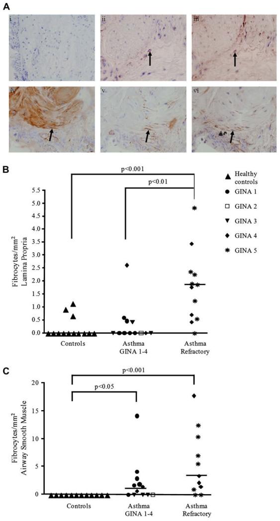
Results: Fifty-one subjects with asthma and 33 control subjects were studied. In bronchial biopsy specimens, the number of fibrocytes was increased in the lamina propria of subjects with severe refractory asthma (median [interquartile range] number, 1.9/mm(2) [1.7/mm(2)]) versus healthy control subjects (median [interquartile range] number, 0/mm(2) [0.3/mm(2)], P < .0001) and in the ASM bundle of subjects with asthma of all severities (subjects with severe asthma, median [interquartile range] number, 3.8/mm(2) [9.4/mm(2)]; subjects with mild-to-moderate asthma, median [interquartile range] number, 1.1/mm(2) [2.4/mm(2)]); healthy control subjects, (median [interquartile range] number, 0/mm(2) [0/mm(2)]); P = .0004). In the peripheral blood the fibrocyte number was also increased in subjects with severe refractory asthma (median [interquartile range] number, 1.4 x 10(4)/mL [2.6 x 10(4)/mL]) versus healthy control subjects (median [interquartile range] number, 0.4 x 10(4)/mL [1.0 x 10(4)/mL], P = .002). We identified that in vitro ASM promotes fibrocyte chemotaxis and chemokinesis (distance of migration after 4.5 hours, 31 microm [2.9 microm] vs 17 microm [2.4 microm], P = .0001), which was in part mediated by platelet-derived growth factor (mean inhibition by neutralizing antibody, 16% [95% CI, 2% to 32%], P = .03) but not by activation of chemokine receptors.
Conclusion: This study provides the first evidence that fibrocytes are present in the ASM compartment in asthma and that ASM can augment fibrocyte migration. The importance of fibrocytes in the development of ASM hyperplasia and airway dysfunction in asthma remains to be determined.
Figures


Similar articles
-
CCL2 release by airway smooth muscle is increased in asthma and promotes fibrocyte migration.Allergy. 2014 Sep;69(9):1189-97. doi: 10.1111/all.12444. Epub 2014 Jun 16. Allergy. 2014. PMID: 24931417 Free PMC article.
-
Fibrocyte localisation to the ASM bundle in asthma: bidirectional effects on cell phenotype and behaviour.Clin Transl Immunology. 2020 Nov 13;9(11):e1205. doi: 10.1002/cti2.1205. eCollection 2020. Clin Transl Immunology. 2020. PMID: 33209301 Free PMC article.
-
Airway smooth muscle and mast cell-derived CC chemokine ligand 19 mediate airway smooth muscle migration in asthma.Am J Respir Crit Care Med. 2006 Dec 1;174(11):1179-88. doi: 10.1164/rccm.200603-394OC. Epub 2006 Sep 7. Am J Respir Crit Care Med. 2006. PMID: 16959919
-
Airway remodeling: potential contributions of subepithelial fibrosis and airway smooth muscle hypertrophy/hyperplasia to airway narrowing in asthma.Allergy Asthma Proc. 1998 Nov-Dec;19(6):353-8. doi: 10.2500/108854198778612672. Allergy Asthma Proc. 1998. PMID: 9876774 Review.
-
Asthma: Pharmacological degradation of the airway smooth muscle layer.Int J Biochem Cell Biol. 2020 Sep;126:105818. doi: 10.1016/j.biocel.2020.105818. Epub 2020 Jul 22. Int J Biochem Cell Biol. 2020. PMID: 32707120 Review.
Cited by
-
IL-17A/F modulates fibrocyte functions in cooperation with CD40-mediated signaling.Inflammation. 2013 Aug;36(4):830-8. doi: 10.1007/s10753-013-9609-z. Inflammation. 2013. PMID: 23400328
-
Extracellular matrix remodelling properties of human fibrocytes.J Cell Mol Med. 2012 Mar;16(3):483-95. doi: 10.1111/j.1582-4934.2011.01344.x. J Cell Mol Med. 2012. PMID: 21595824 Free PMC article.
-
Fibrocytes in the Pathogenesis of Chronic Fibrotic Lung Disease.Curr Respir Med Rev. 2013 Feb;9(1):34-41. doi: 10.2174/1573398x11309010005. Curr Respir Med Rev. 2013. PMID: 27512347 Free PMC article.
-
Mesenchymal stem cells regulate airway contractile tissue remodeling in murine experimental asthma.Allergy. 2014 Jun;69(6):730-40. doi: 10.1111/all.12392. Epub 2014 Apr 21. Allergy. 2014. PMID: 24750069 Free PMC article.
-
Reduced suppressive effect of β2-adrenoceptor agonist on fibrocyte function in severe asthma.Respir Res. 2017 Nov 21;18(1):194. doi: 10.1186/s12931-017-0678-7. Respir Res. 2017. PMID: 29162108 Free PMC article.
References
-
- Global Initiative for Asthma [Accessed September 17, 2008];Global Initiative for Asthma guidelines. www.ginasthma.com
-
- Chanez P, Wenzel SE, Anderson GP, Anto JM, Bel EH, Boulet L, et al. Severe asthma: what are the important questions? J Allergy Clin Immunol. 2007;119:1337–48. - PubMed
-
- Proceedings of the American Thoracic Society Workshop on Refractory Asthma. Current understanding, recommendations, and unanswered questions. Am J Respir Crit Care Med. 2000;162:2341–51. - PubMed
-
- Wardlaw AJ, Brightling C, Green R, Woltmann G, Pavord I. Eosinophils in asthma and other allergic diseases. Br Med Bull. 2000;56:985–1003. - PubMed
-
- Brightling CE, Bradding P, Symon FA, Holgate ST, Wardlaw AJ, Pavord ID. Mast-cell infiltration of airway smooth muscle in asthma. N Engl J Med. 2002;346:1699–705. - PubMed
Publication types
MeSH terms
Substances
Grants and funding
LinkOut - more resources
Full Text Sources
Other Literature Sources
Medical
