

Nasal cavity changes and the respiratory standard after maxillary expansion
- PMID: 19082360
- PMCID: PMC9445931
- DOI: 10.1016/S1808-8694(15)31388-4
Nasal cavity changes and the respiratory standard after maxillary expansion
Abstract
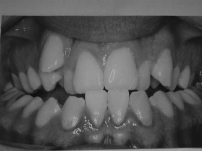
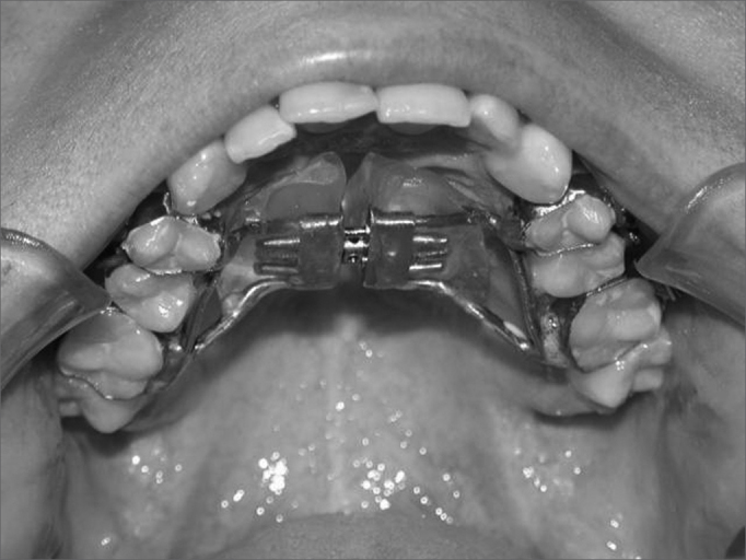
Mandibular cross-sectional deficiency is a dentofacial defect in connection with the narrowing of the mandibular arch width. This abnormality is a significant etiopathogenic factor and it is often associated with nasal breathing difficulties. This atresia may be treated through Rapid Maxillary Expansion or Surgically Assisted Rapid Maxillary Expansion, depending on the patient's age. Both procedures will change the craniofacial structure, especially the nasal cavity.
Aim: Based on literature review, the purpose of this paper was to report the relationship among maxillary expansion, nasal cavity and Nasal Airflow Resistance.
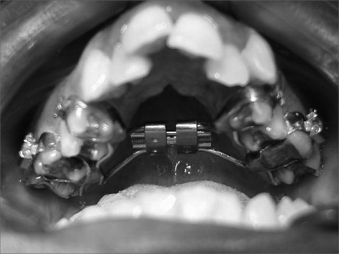
Method: A non-systematic literary review was conducted in search of experimental studies to treat maxillary atresia. Papers considering Rapid Maxillary Expansion and Surgically Assisted Rapid Maxillary Expansion were included, whereas those using Maxillary Expansion through Segmented Osteotomy were excluded.
Result: Rapid Maxillary Expansion and Surgically Assisted Rapid Maxillary Expansion cause dentofacial changes, especially in the nasal cavity. Consequently, the nose width enlarges, reducing Nasal Airflow Resistance.
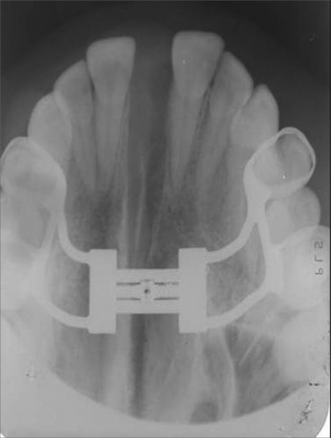
Conclusion: Anteroposterior cephalometric studies show evidence of an enlarged nasal cavity following maxillary expansion.
Figures
References
-
- Haas AJ. Rapid Expansion of the maxillary dental arch and nasal cavity by opening the midpalatal suture. Angle Orthod. 1961;31(2):73–90.
-
- Wertz RA. Changes in nasal airflow incident to rapid maxillary expansion. Angle Orthod. 1968;33(1):1–11. - PubMed
-
- Manganello LC, Cappellette M. Tratamento cirúrgico de pacientes com palato ogival e com obstrução nasal. Rev Assoc Paul Cir Dent. 1996;50(1):79–81.
-
- Andrade A, Oliveira LCS. Avaliação de deformidade septal por vide-ofibroscopia nasal em pacientes adultos com atresia transversal de maxila. Rev Bras Otorrinolaringol. 2002;68(5):1–11.
-
- Hartgerink DV, Vig PS, Abbott DW. The effect of rapid maxillary expansion an nasal airway resistance. Am J Orthod Dentofacial Orthop. 1987;92(5):381–389. - PubMed
Uncited Reference
-
- Trevisan RA. In: Cirurgia Ortognática. Araújo A, editor. Editora Santos; São Paulo: 1999. Expansão rápida do palato: ortodontia × cirurgia; pp. 213–222.
Publication types
MeSH terms
LinkOut - more resources
Full Text Sources
