. 2009 Aug;64(3):433-43.
doi: 10.1007/s00280-008-0888-2. Epub 2008 Dec 12.

Heat shock protein 90 inhibition abrogates hepatocellular cancer growth through cdc2-mediated G2/M cell cycle arrest and apoptosis

Affiliations

Heat shock protein 90 inhibition abrogates hepatocellular cancer growth through cdc2-mediated G2/M cell cycle arrest and apoptosis

Go Watanabe et al. Cancer Chemother Pharmacol. 2009 Aug.

Abstract

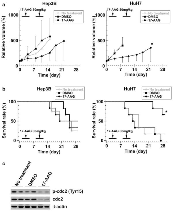
Purpose: 17-(demethoxy), 17-allylamino geldanamycin (17-AAG) suppresses growth in some cancers by inhibiting Heat shock protein 90 (Hsp90). We examined the effects of 17-AAG-mediated Hsp90 inhibition on human hepatocellular carcinoma (HCC) growth in vitro and in vivo.

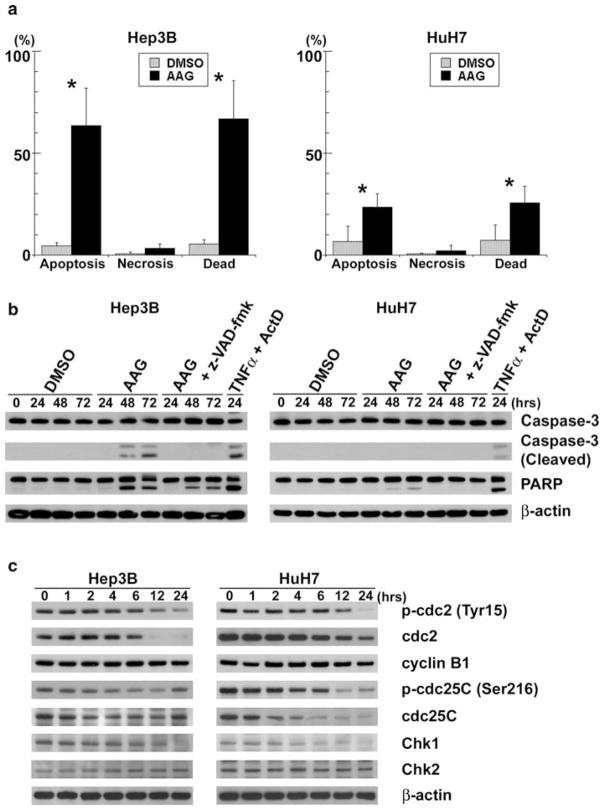
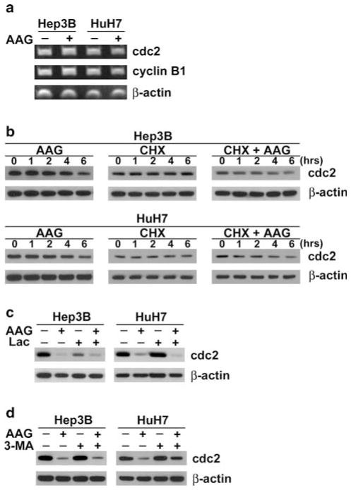
Methods: Human HCC cell lines, Hep3B and HuH7, were exposed to 17-AAG and cell viabilities and apoptosis were determined. Cell cycle profiles were analyzed and the G(2)/M cell cycle checkpoint proteins cdc2 and cyclin B1 were examined. Studies were performed to determine whether 17-AAG-mediated cdc2 decrease was due to altered gene expression, transcription, or protein degradation. The effects of 17-AAG on Hep3B and HuH7 xenograft growth in athymic nude mice were also examined.

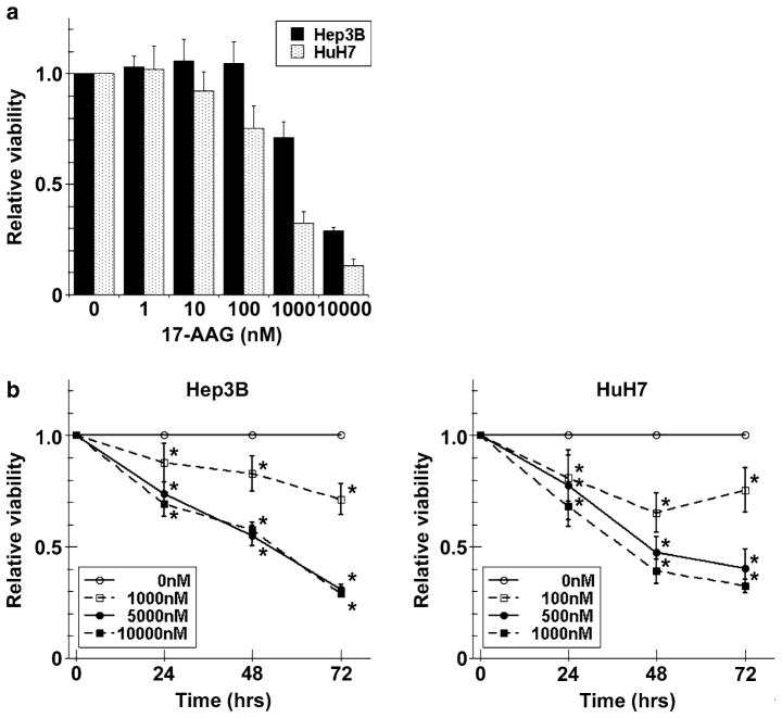
Results: Hep3B and HuH7 treated with 17-AAG versus untreated controls showed decreased cell viability and increased apoptosis. Cells treated with 17-AAG also showed an increased fraction in G(2)/M phase and an associated decrease in cdc2 through protein degradation rather than through other mechanisms. Hsp90 inhibition by 17-AAG also decreased HCC xenograft growth in association with decreased cdc2 expression.

Conclusions: 17-AAG-mediated inhibition of Hsp90 abrogates human HCC cell growth in vitro and in vivo through cdc2 decrease, which in turn induces G(2)/M cell cycle arrest and apoptosis. Hsp90 is a mediator of HCC growth and survival and its inhibition may serve as a potential treatment.


Figures

Fig. 1
Fig. 1
Viability effects of 17-AAG on human HCC cells. a Hep3B and HuH7 cells were incubated with a range of 17-AAG concentrations for 72 h, viabilities were measured by MTT assay and expressed as relative viabilities to DMSO control cells. b Hep3B and HuH7 cells were incubated with or without a range of 17-AAG concentrations (including IC50) for 24, 48, and 72 h, then viabilities were measured by MTT assay. The results are shown as relative cell viability as compared to controls (DMSO alone) at various concentrations of 17-AAG for up to 72 h and the values are expressed as the mean ± SD of three independent experiments * P < 0.05
Fig. 2
Fig. 2
17-AAG induces apoptosis and decreases the expression of cell cycle-related protein. a Apoptotic and necrotic cells were counted by fluorescence microscopy of Hep3B and HuH7 cells after live cell staining with Hoechst 33342 and PI. Dead cells were the sum of apoptotic and necrotic cells. Each of the cell counts were expressed as a percentage of the total cell number, and significant differences in percent cell number between 17-AAG treated and untreated groups were indicated (* P < 0.05). b Hep3B and HuH7 cells were treated with 17-AAG (AAG) at respective IC50, 20 μM z-VAD-fmk or vehicle alone (DMSO) for up to 72 h. Cell lysates were analyzed by immunoblots for uncleaved and cleaved caspase-3, PARP and β-actin. As a positive control, the cells treated with 20 ng/ml TNF-α (TNFα) and 200 ng/ml Actinomycin D (ActD) for 24 h were used. c Hep3B and HuH7 cells were incubated with 17-AAG at respective IC50 for up to 24 h. Protein extracts were then analyzed by immunoblots for cdc2 (total and phosphorylated at Tyr15), cyclin B1, cdc25C (total and phosphorylated at Ser216), Chk1, Chk2 and β-actin
Fig. 3
Fig. 3
17-AAG mediates cdc2 decrease. a Hep3B and HuH7 cells were incubated with 17-AAG (AAG) (+) at respective IC50 concentrations or DMSO vehicle alone (−) for 24 h. Total mRNA were extracted then analyzed by RT-PCR for cdc2, cyclin B1 and β-actin mRNA. b Protein cell lysates were extracted from Hep3B and HuH7 cells treated for up to 6 h with 10 μg/ml of cycloheximide (CHX) in the presence or absence of respective IC50 concentration of 17-AAG (AAG), and then analyzed by immunoblots for cdc2 and β-actin. c Protein cell extracts were derived from HCC cells incubated for 24 h in the presence or absence of IC50 of 17-AAG (AAG) and/or 10 μM lactacystin (Lac). Immunoblots were probed for cdc2 and β-actin. d Each cell line was exposed for 24 h with DMSO (control), IC50 of 17-AAG (AAG), with or without 3-methyladenine (3-MA) at 10 mM. Cell lysates were analyzed by immunoblots for cdc2 and β-actin
Fig. 4
Fig. 4
Inhibition of cdc2 induces apoptosis. Cell lysates of Hep3B and HuH7 cells treated with 50 μM Roscovitine (Ros), 20 μM z-VAD-fmk or vehicle alone (DMSO) for up to 72 h were analyzed by immunoblots for uncleaved and cleaved caspase-3, PARP, total and phosphorylated (Tyr15) cdc2, and β-actin. As a positive control, the cells treated with 20 ng/ml TNF-α (TNFα) and 200 ng/ml Actinomycin D (ActD) for 24 h were used
Fig. 5
Fig. 5
17-AAG decreases human HCC xenograft growth. a Once Hep3B and HuH7 xenografts reached 100 mm3 (day 0), each nude mouse received 17-AAG (80 mg/kg/day, days 1–4 and 8–11), DMSO (vehicle only, same days) or no treatment (n = 6 per group). The effect of 17-AAG on HCC growth was determined by measuring Hep3B and HuH7 xenografts dimensions, calculating tumor volumes and expressing these as a percentage of the day 0 volumes (relative tumor volume). The values are shown as the mean ± SD (* P < 0.05, vs. DMSO). b Each mouse was euthanized when the largest tumor diameter reached 15 mm, and this event was defined as “tumor-related death”. Tumor-related survival for mice with Hep3B and HuH7 xenografts were determined by Kaplan-Meier analysis, and log rank test was used to detect any differences between the three treatment groups. (* P < 0.05, vs. DMSO) c Protein extracts from HuH7 cell xenografts at day 14 were analyzed by immunoblots for cdc2 (total and phosphorylated at Tyr15) and β-actin

Similar articles

Cited by

References

    1. Bosch FX, Ribes J, Diaz M, et al. Primary liver cancer: worldwide incidence and trends. Gastroenterology. 2004;127:S5–S16. - PubMed
    1. Young JC, Moarefi I, Hartl FU. Hsp90: a specialized but essential protein-folding tool. J Cell Biol. 2001;154:267–273. - PMC - PubMed
    1. Welch WJ, Feramisco JR. Purification of the major mammalian heat shock proteins. J Biol Chem. 1982;257:14949–14959. - PubMed
    1. Luk JM, Lam CT, Siu AF, et al. Proteomic profiling of hepatocellular carcinoma in Chinese cohort reveals heat-shock proteins (Hsp27, Hsp70, GRP78) up-regulation and their associated prognostic values. Proteomics. 2006;6:1049–1057. - PubMed
    1. Calvisi DF, Pascale RM, Feo F. Dissection of signal transduction pathways as a tool for the development of targeted therapies of hepatocellular carcinoma. Rev Recent Clin Trials. 2007;2:217–236. - PubMed

MeSH terms