. 2009 Jun;34(6):1138-49.
doi: 10.1007/s11064-008-9886-3. Epub 2008 Dec 12.

Immediate and delayed consequences of xanomeline wash-resistant binding at the M3 muscarinic receptor

Affiliations

Immediate and delayed consequences of xanomeline wash-resistant binding at the M3 muscarinic receptor

Meredith J Noetzel et al. Neurochem Res. 2009 Jun.

Abstract

Xanomeline is thought to be a M1/M4 functionally selective agonist at muscarinic receptors. We have previously demonstrated that it binds in a unique manner at the M1 receptor. In the current study, we examined the ability of xanomeline to bind to the M3 receptor and determined the long-term consequences of this mode of binding in Chinese hamster ovary cells expressing M3 receptors. Xanomeline binds in a reversible and wash-resistant manner at the M3 receptor and elicits a functional response under both conditions. Long-term exposure to xanomeline resulted in changes in the binding profile of [(3)H]NMS and a decrease in cell-surface receptor density. Additionally, pretreatment with xanomeline was associated with antagonism of the functional response to subsequent stimulation by conventional agonists. Our results indicate that xanomeline binds to and activates the M3 muscarinic receptor in a wash-resistant manner, and that this type of binding results in time-dependent receptor regulation.


Figures

Fig. 1
Fig. 1
Time course of the development of xanomeline wash-resistant binding in CHO cells stably expressing human M3 muscarinic receptors. Cells were incubated with xanomeline for increasing lengths of time followed by extensive washing and determination of 0.2 nM [3H]NMS specific binding. Values represent the means ± SE of three experiments conducted in triplicate. Binding in the absence of xanomeline pretreatment is denoted by 100%. Individual experiments were normalized to control binding in the absence of xanomeline treatment, which had a mean of 5,800 dpm/well
Fig. 2
Fig. 2
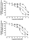
Inhibition of specific [3H]NMS binding following treatment with increasing concentrations of xanomeline in CHO cells expressing the human M3 receptor. The binding of 0.2 nM [3H]NMS was determined in the continuous presence of increasing concentrations of xanomeline (closed squares). Cells were treated with xanomeline for 1 min (closed circles, a) or 1 h (closed triangles, b) followed by washing and immediate application in the binding assay or followed by washing and waiting for 24 h (open circles, A) or 23 h (open triangles, b), respectively. An additional group of cells was treated with xanomeline for 24 h followed by washing and use in binding assay (closed diamonds). Following all pretreatments cells were incubated with 0.2 nM [3H]NMS for 1 h at 37°C. Individual experiments were normalized to control binding in the absence of xanomeline that averaged 21,300 dpm/well. Values represent the means ± SE. of 3–8 experiments conducted in triplicate
Fig. 3
Fig. 3
Effects of xanomeline pretreatment on [3H]NMS saturation binding in CHO cells expressing human M3 receptors. Cells were pretreated with 10 μM xanomeline for 1 h followed by washing and immediate application in the binding assay (closed triangles) or after 23 h incubation in the absence of free xanomeline (open triangles). An additional group of cells was treated with xanomeline for 24 h followed by washing and addition to the binding assay mixture (closed diamonds). Untreated control cells (closed squares) and xanomeline-pretreated and washed cells were subsequently incubated with increasing concentrations of [3H]NMS (0.01–6.5 nM) for 1 h at 37°C. Non-specific binding was determined in the presence of 10 μM atropine. Values represent the means ± SE of four experiments conducted in triplicate
Fig. 4
Fig. 4
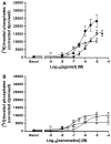
M3 receptor activation by xanomeline reversible and wash-resistant binding. a Cells were incubated at 37°C for 1 h with increasing concentrations of carbachol (closed squares), pilocarpine (closed triangles) or xanomeline (open diamonds) in the presence of 10 mM lithium chloride and inositol phosphate production was determined; b cells were pretreated with increasing concentrations of xanomeline for 1 min (open circles) or 1 h (open diamonds) followed by washing and determination of the accumulation of inositol phosphates in the absence of free xanomeline. In the third group (diamonds) xanomeline was only present during determination of inositol phosphates accumulation. Values represent the means ± SE. of 3–5 experiments conducted in triplicate
Fig. 5
Fig. 5
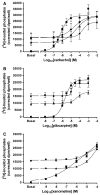
Effects of acute xanomeline exposure on the ability of muscarinic agonists to stimulate the production of inositol phosphates. Cells were pretreated with 10 μM xanomeline for 1 min (closed circles) or 1 h (closed triangles) followed by washing and immediate use. Subsequently, cells were treated with increasing concentrations of a carbachol; b pilocarpine; or c xanomeline and the accumulation of inositol phosphates was determined over a 1 h incubation period at 37°C in the presence of 10 mM lithium chloride for untreated cells (closed squares) and those pretreated with xanomeline. a An additional group of cells was simulated simultaneously with increasing concentrations of carbachol and 10 μM xanomeline (closed diamonds). Values represent the means ± SE. of 3–7 experiments conducted in triplicate
Fig. 6
Fig. 6
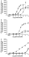
Effects of prolonged xanomeline exposure on the ability of muscarinic agonists to stimulate the production of inositol phosphates. Cells were pretreated with 10 μM xanomeline for 1 min (open circles) or 1 h (open triangles) followed by washing and waiting 24 or 23 h, respectively. An additional group of cells was treated with xanomeline for 24 h (closed diamonds) prior to washing. Subsequently, cells were treated with increasing concentrations of a carbachol; b pilocarpine; or c xanomeline and the accumulation of inositol phosphates was determined over a 1 h incubation period at 37°C in the presence of 10 mM lithium chloride for untreated cells (closed squares) or those pretreated with xanomeline. Values represent the means ± SE. of 3–7 experiments conducted in triplicate
Fig. 7
Fig. 7
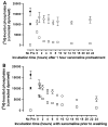
Time course of switching of the pharmacological profile of xanomeline from an agonist to an antagonist. a Cells were pretreated with 10 μM xanomeline for 1 h followed by washing and waiting for increasing lengths of time (0–23 h); b Cells were pretreated with 10 μM xanomeline for increasing amounts of time (30 min–24 h) prior to washing. Subsequently, the accumulation of inositol phosphates was determined over a 1 h incubation period in the presence of 10 mM lithium chloride following no further stimulation (open squares) or stimulation with 10 μM carbachol (open circles). For comparison, untreated cells were stimulated with carbachol (closed circles) or xanomeline (closed diamonds). No pre: untreated cells that were not exposed to xanomeline. Values represent the means of two experiments conducted in triplicate

Similar articles

Cited by

References

    1. Caulfield MP, Birdsall NJ. International union of pharmacology Xvii. Classification of muscarinic acetylcholine receptors. Pharmacol Rev. 1998;50:279–290. - PubMed
    1. Wess J. Muscarinic acetylcholine receptor knockout mice: novel phenotypes and clinical implications. Annu Rev Pharmacol Toxicol. 2004;44:423–450. 10.1146/annurev.pharmtox.44.101802. 121622. - PubMed
    1. Eglen RM. Muscarinic receptor subtype pharmacology and physiology. Prog Med Chem. 2005;43:105–136. doi: 10.1016/S0079-6468(05)43004-0. - DOI - PubMed
    1. Volpicelli LA, Levey AI. Muscarinic acetylcholine receptor subtypes in cerebral cortex and hippocampus. Prog Brain Res. 2004;145:59–66. doi: 10.1016/S0079-6123(03)45003-6. - DOI - PubMed
    1. Francis PT, Palmer AM, Snape M, et al. The cholinergic hypothesis of alzheimer’s disease: a review of progress. J Neurol Neurosurg Psychiatry. 1999;66:137–147. - PMC - PubMed

Publication types