

Review
doi: 10.1016/j.jbmt.2008.03.004.
Epub 2008 May 21.
Epi-perineurial anatomy, innervation, and axonal nociceptive mechanisms
Affiliations
- PMID: 19083672
- PMCID: PMC2610338
- DOI: 10.1016/j.jbmt.2008.03.004
Item in Clipboard
Review
Epi-perineurial anatomy, innervation, and axonal nociceptive mechanisms
J Bodyw Mov Ther.
2008 Jul.
Abstract
Nerves are usually viewed as simple conduits of electrical signals to make muscles move and enable sensation. However, recent data have shown that the axons within nerves are capable of responding to their environment. Nerves have a very specialized anatomy and physiology, and are capable of mediating certain types of pain. This synopsis introduces the reader to these concepts, which can be incorporated into clinical decision-making.
Figures
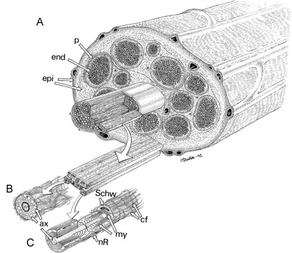
Peripheral nerve anatomy. A. Endoneurium-covered axons (end) are bundled by perineurium (p) into fascicles. These fascicles are in turn covered by epineurium (epi). B, C: Specializations of the Schwann cells that surround unmyelinated (B) and myelinated (C) axons. Numerous unmyelinated axons (ax) are embedded by invagination into the cytoplasm of a single Schwann cell (Sch). Each myelinated axon has multiple layers of Schwann cell membrane wrapped around it, forming myelin (my), interrupted at regular intervals by nodes of Ranvier (nR). (From Lundborg G 1988, used with permission.)
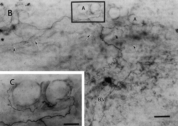
Peripherin-like immunostaining in nervi nervorum and vasa nervorum of rat sciatic nerve (whole mount). a. Montage demonstrating nerves following a blood vessel (BV, outlined by the fibers flanking the label) and leaving it to terminate in epineurium (arrows) and associated with adipose tissue (A). Note extensive branching. b, enlargement of area marked in a, detailing adipose innervation. M = mast cell, A = adipocyte. Scale bars in a = 50 μm, b = 20 μm. (From Bove GM & Light AR 1995a, used with permission.)
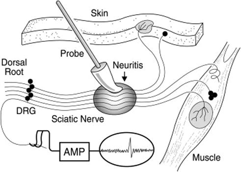
Axonal mechanical sensitivity is induced by nerve inflammation. The sciatic nerve is depicted in the middle of the image, with the shaded oval area indicating CFA-induced inflammation. The individual neurons lead to receptive fields in muscle or skin (right and top), and centrally to dorsal roots (left), for recording. When inflamed, the normally insensitive axons become mechanically sensitive. DRG = dorsal root ganglion. (From Bove GM et al 2003, used with permission.)
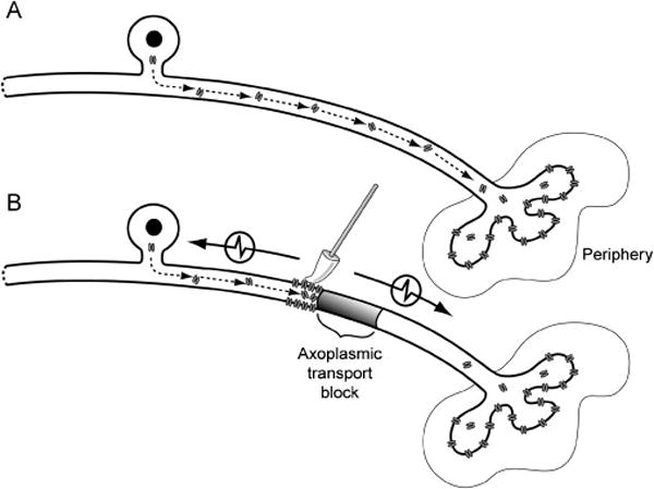
A. Components required for mechanical sensitivity are transported from the cell body of a single C-fiber neuron to the periphery for insertion at the terminals. B. Blocking axoplasmic transport leads to the accumulation and insertion of mechanosensitive components proximal to the site of axoplasmic blockade. Mechanical stimulation of the axon membrane therefore becomes an effective stimulus to generate action potentials that pass in both directions along the axon (denoted by arrows). (From Dilley A & Bove GM 2008, used with permission.)
References
-
- Armstrong BD, et al. Induction of neuropeptide gene expression and blockade of retrograde transport in facial motor neurons following local peripheral nerve inflammation in severe combined immunodeficiency and BALB/C mice. Neuroscience. 2004;129:93–99. - PubMed
-
- Asbury AK, Fields HL. Pain due to peripheral nerve damage: an hypothesis. Neurology. 1984;34:1587–1590. - PubMed
-
- Bove GM, Light AR. Calcitonin gene-related peptide and peripherin immunoreactivity in nerve sheaths. Somatosensory and Motor Research. 1995a;12:49–57. - PubMed
-
- Bove GM, Light AR. Unmyelinated nociceptors of rat paraspinal tissues. Journal of Neurophysiology. 1995b;73:1752–1762. - PubMed
-
- Bove GM, et al. Inflammation induces ectopic mechanical sensitivity in axons of nociceptors innervating deep tissues. Journal of Neurophysiology. 2003;90:1949–1955. - PubMed
Publication types
MeSH terms
Grants and funding
LinkOut - more resources
Full Text Sources
Other Literature Sources
Medical
