. 2009 Mar;88(3):512-21.
doi: 10.1016/j.exer.2008.11.013. Epub 2008 Dec 3.

The role of Akt/protein kinase B subtypes in retinal ischemic preconditioning

Affiliations

The role of Akt/protein kinase B subtypes in retinal ischemic preconditioning

John C Dreixler et al. Exp Eye Res. 2009 Mar.

Abstract

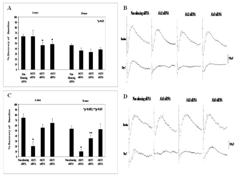
Potent endogenous protection from ischemia can be induced in the retina by ischemic preconditioning (IPC). Protein kinase B/Akt is a cellular survival factor. We hypothesized that Akt was integral to IPC based upon differential effects of Akt subtypes. Rats were subjected to retinal ischemia after IPC or IPC-mimicking by the opening of mitochondrial KATP (mKATP) channels. The effects of blocking Akt using wortmannin, API-2, or small interfering RNA (siRNA) were examined. Electroretinography assessed functional recovery after ischemia, and TUNEL examined retinal ganglion cell apoptosis. We studied the relationship between Akt activation and known initiators of IPC, including adenosine receptor stimulation and the opening of mKATP channels. The PI-3 kinase inhibitor wortmannin 1 or 4 mg/kg (i.p.), the specific Akt inhibitor API-2, 5-500 microM in the vitreous, or intravitreal siRNA directed against Akt2 or -3, but not Akt1, significantly attenuated the neuroprotective effect of IPC. Interfering RNA against any of the three Akt subtypes significantly but time-dependently attenuated mKATP channel opening to mimic IPC. Adenosine A1 receptor blockade (DPCPX), A2a blockade (CSC), or the mKATP channel blocker 5-hydroxydecanoic acid significantly attenuated Akt activation after IPC. Interfering RNA directed against Akt subtypes prevented the ameliorative effect of IPC on post-ischemic apoptosis. All three Akt subtypes are involved in functional retinal neuroprotection by IPC or IPC-mimicking. Akt is downstream of adenosine A1 and A2a receptors and mKATP channel opening. The results indicate the presence in the retina of robust and redundant endogenous neuroprotection based upon subtypes of Akt.


Conflict of interest statement

Proprietary Interest: None

Figures

Fig. 1
Fig. 1
Inhibition of Akt activation by API-2 attenuated recovery of the electroretinogram (ERG) a- and b-waves after preconditioning (IPC) followed by ischemia. Eyes were subjected to IPC, and ischemia followed 24 h later. A. Both a- and b-wave recovery after ischemia in the preconditioned rat retina were significantly diminished in a dose-dependent manner by intravitreal injection of the Akt phosphorylation inhibitor API-2. Vehicle control was 200 μM HCl (in PBS). B. Representative ERG traces for the baseline and 7 days after ischemia. C. Western blot quantification of phosphorylated Akt showed that API-2 significantly inhibited Akt activation 1 h after API-2 intravitreal injection. D. Representative Western blot bands of the 60 kD phosphorylated Akt, with no change in levels of total Akt, indicating equal loading of protein.
Fig. 2
Fig. 2
PI-3K inhibition, by wortmannin, blocked neuroprotection with ischemic preconditioning (IPC). Eyes were subjected to IPC, and ischemia followed 24 h later. A. Representative traces of the electroretinogram (ERG) for baseline and 7 days after ischemia. B. IPC recovery shown on ERG (both a- and b-wave) in the rat retina was significantly diminished by wortmannin.
Fig. 3
Fig. 3
Akt subtype siRNA specificity. A. Akt3 protein levels were significantly reduced 24 h after intravitreal injection as compared to the non-silencing siRNA control. Representative Western blots are shown (right) as well as rhodopsin protein levels that serve as a control. B. Western blot analysis of Akt1 siRNA show decreases in their respective Akt subtype protein levels 54 h after injection. Actin served as a control.
Fig. 4
Fig. 4
Immunostained cryosections of retina injected with Akt2 siRNA showed significant reduction of Akt2 levels in the retinal ganglion cell (RGC) and inner nuclear layers (INL). Akt2 levels were unchanged in the photoreceptor (PR) layer (Fig. 5A). Representative immunostaining of Akt2 is shown (Fig. 5B).Retinal cryosections from paired eyes injected with siRNA to Akt2 or non-silencing siRNA and harvested 24 h later were reacted with anti-Akt2 antibody and fluorescein-conjugated secondary antibody. Green color indicates staining for Akt2 protein (shown in the middle panels). DAPI (far left images) are for orientation (cell layers shown in white letters), where cell nuclei stain in blue. The right panels show double labeling for DAPI and Akt2. White arrows indicate some of the cells positive for Akt2 protein in the GCL and INL; also shown in the far right panel are cells double labeling for DAPI and Akt2, indicating cellular localization of Akt2 protein in cells in the RGC and INL layers. It is evident that Akt2 positivity was diminished by siRNA directed against it. Fig. 5C is a section reacted with non-immune serum to indicate levels of background staining.
Fig. 5
Fig. 5
Akt subtype specific siRNA blocked neuroprotection with ischemic preconditioning (IPC). Eyes were subjected to IPC and ischemia followed 24 h later. A. Representative traces of the electroretinogram (ERG) at baseline and 7 days after ischemia. B. Injection of 2 μl of 3 μM siRNA to Akt2 or Akt3 into the vitreous 6 h before IPC significantly attenuated post-ischemic recovery of both the a- and b-waves on ERG. Akt1 siRNA was ineffective. Intravitreal of injections of a non-silencing siRNA served as the control. C. Akt2 siRNA intravitreal injection before sham IPC, followed by ischemia 24 h later, did not alter the recovery compared to Akt2 siRNA with IPC and ischemia.
Fig. 6
Fig. 6
Akt subtype specific siRNA attenuated ischemic preconditioning (IPC)-mimicking by diazoxide in a time-dependent manner. Rats were injected with diazoxide, and ischemia followed 24 h later. A. Intravitreal injection of 2 μl of 3 μM subtype-specific Akt2 and Akt3 siRNA 6 h before diazoxide injection (i.p.) significantly decreased IPC-mimicking by the mKATP channel activator for the ERG a-wave. Akt1 siRNA was ineffective. A non-silencing siRNA served as control. B. Representative traces of electroretinogram (ERG) at baseline and 7 days after ischemia for the 6 h prior to diazoxide-injected groups. C. Akt1 siRNA significantly reduced the post-ischemic recovery (for both a- and b-waves) when injected 24 h before diazoxide. Akt2 siRNA significantly attenuated the recovery of b-wave on ERG. Akt3 siRNA was without effect. D. Representative ERG traces for the baseline and 7 days after ischemia for the 24 h experiments.
Fig. 7
Fig. 7
Akt activation was reduced by mKATP channel and adenosine receptor antagonists. A. Western blot analysis of phosphorylated Akt protein levels were significantly reduced compared to levels at 6 h after ischemic preconditioning (IPC), by the specific mKATP channel blocker 5-HD at a concentration of 200 mg/kg, the adenosine A1 receptor antagonist DPCPX and the adenosine A2 receptor inhibitor CSC. These results suggest that Akt activation is downstream from both the mKATP channel and adenosine receptor pathways. B. Representative Western blot bands of the 60 kD phosphorylated Akt, with no change in the beta tubulin control in the paired eyes.
Fig. 8
Fig. 8
Fluorescent TUNEL staining and Akt subtype involvement in apoptosis. Shows representative images of the fluorescent TUNEL staining for the normal RGC layer (left) versus the ischemic (right). Images are shown with altered/false colors where the arrows denote co-localization of the fluorescent TUNEL stain (red) and the DAPI (green).

References

    1. Alessi DR, Andjelkovic M, Caudwell B, Cron P, Morrice N, Cohen P, Hemmings BA. Mechanism of activation of protein kinase B by insulin and IGF-1. EMBO J. 1996;15:6541–6551. - PMC - PubMed
    1. Cardone MH, Roy N, Stennicke HR, Salvasen GS, Franke TF, Stanbridge E, Frisch S, Reed JC. Regulation of cell death protease caspase-9 by phosphorylation. Science. 1998;282:1318–1321. - PubMed
    1. Chavarria T, Valenciano AI, Mayordomo R, Egea J, Comella JX, Hallbook F, de Pablo F, de la Rosa EJ. Differential, age-dependent MEK-ERK and PI3K-Akt activation by insulin acting as a survival factor during embryonic retinal development. Dev Neurobiol. 2007;67:1777–1788. - PubMed
    1. Chong ZZ, Li F, Maiese K. Oxidative stress in the brain: novel cellular targets that govern survival during neurodegenerative disease. Prog Neurobiol. 2005;75:207–246. - PubMed
    1. Datta SR, Dudek H, Tao X, Masters S, Fu H, Gotah Y, Greenberg ME. Akt phosphorylation of BAD couples survival signals to the cell-intrinsic death machinery. Cell. 1997;911:231–241. - PubMed

Publication types

MeSH terms