. 2009 Mar 4;134(2):132-40.
doi: 10.1016/j.jconrel.2008.10.020. Epub 2008 Nov 12.

Polymersome delivery of siRNA and antisense oligonucleotides

Affiliations

Polymersome delivery of siRNA and antisense oligonucleotides

Younghoon Kim et al. J Control Release. .

Erratum in

  • J Control Release. 2009 Aug 4;137(3):256. Williams, Jason [corrected to Williams, Jason H]; Sirsi, Shashank, [corrected to Sirsi, Shashank R]; Lutz, Gordon [corrected to Lutz, Gordon J]

Abstract

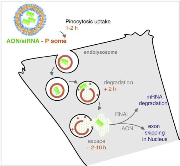
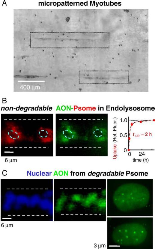
siRNA and antisense oligonucleotides, AON, have similar size and negative charge and are often packaged for in vitro delivery with cationic lipids or polymers-but exposed positive charge is problematic in vivo. Here we demonstrate loading and functional delivery of RNAi and AON with non-ionic, nano-transforming polymersomes. These degradable carriers are taken up passively by cultured cells after which the vesicles transform into micelles that allow endolysosomal escape and delivery of either siRNA into cytosol for mRNA knockdown or else AON into the nucleus for exon skipping within pre-mRNA. Polymersome-mediated knockdown appears as efficient as common cationic-lipid transfection and about half as effective as Lenti-virus after sustained selection. For AON, initial results also show that intramuscular injection into a mouse model of muscular dystrophy leads to the expected protein expression, which occurs along the entire length of muscle. The lack of cationic groups in antisense polymersomes together with initial tests of efficacy suggests broader utility of these non-viral carriers.


Figures

Fig. 1
Fig. 1
Cell uptake and endolysosomal escape of polymersome encapsulated oligonucleotides. Based on recent studies (Ahmed et al. [25]), polymer vesicles of ∼100 nm are taken up non-specifically via pinocytosis over several hours. Controlled release polymer vesicles degrade in the low pH lumen of the lysosome, releasing their payload, and eventually lysing the confining lipid bilayer. The released payload is then free to diffuse through the cell.
Fig. 2
Fig. 2
Encapsulation, degradation, release, and lysis of antisense with controlled release polymersomes (Psome). (A) Fluorescence microscope images of AON (green) loaded in biodegradable OCL-based Psome (red, due to fluorescent copolymer) after formation of large unilamellar vesicles. Inset shows edge-bright intensity profile for Psome membrane and the filled lumen profile for AON, indicating a lack of membrane interaction. (B) Fluorescence images of siRNA (green) loaded into fluorescently-labeled OLA nano-polymersomes (red). (C) Transition from vesicles to micelles occurs over tens of hours as 100 nm vesicles are lost and 40 nm micelles emerge. The indicated fit of the decay has a t = 3 h offset to account for stable 100−110 nm vesicles, and then a micellization time constant of t = 2 h. (D) Release kinetics from self-porating vesicles are consistent with degradation kinetics, and increase with increasing temperature; at 4 °C, leakage of oligo is undetectable for days. Release is accelerated under acidic conditions, typical of endolysosomes inside cells. (E) Degradable polymersomes are membrane lytic at concentrations of ∼100 mg/ml, which are attainable in endolysosomes containing one or more polymersomes. Red cell hemolysis is the standard assay used. TX 100 is the non-ionic detergent triton X-100 at just 10 mg/ml and shown here to rapidly lyse membranes compared to lysis times for 100 mg/ml copolymers of 10−20 h. (For interpretation of the references to color in this figure legend, the reader is referred to the web version of this article.)
Fig. 3
Fig. 3
Delivery of siRNA and knockdown of lamin-A/C. (A) A549 cancer cells (outlined) take up fluorescently-labeled polymersomes (red, due to PKH26 dye) loaded with siRNA (green) within several hours. (B,C) Polymersome delivery of siRNA induces knockdown of the lamin A/C proteins just as effectively as Lipofectamine 2000 (LF2k). Naked siRNA does not induce measurable changes in lamin A/C expression, while lentiviral delivery followed by antibiotic selection results in about 2-fold greater knockdown.
Fig. 4
Fig. 4
Uptake of degradable and nondegradable polymersomes in micropatterned C2C12 myotubes. (A) Micro patterning of myotubes minimizes cell layering and provides well-defined geometries for studying cellular uptake and localization. (B) Non-degradable PEG-PBD vesicles (red, due to fluorescent copolymer) were taken up by quiescent myotubes over a few hours, and showed a perinuclear localization for several days. (C) Biodegradable OCL-Psomes showed efficient nuclear delivery of fluorescent-AON within 24 h. High magnification images of myonuclei show clear and diffuse nuclear uptake as well as a few punctuate localizations. The diffuse AON is highly mobile, while punctates show little recovery after FRAP (see Fig. S2).
Fig. 5
Fig. 5
Intramuscular AON delivery and Dystrophin expression in mdx mice. (A) Mechanism of action of exon-skipping AON: after endolyosomal escape, AON enters the nucleus and binds target premRNA at spliceosomal recognition sites. This causes the splicosomal machinery to fail at these sites, resulting in an mRNA that lacks the targeted exons which are degraded along with introns. (B) Nuclear delivery of Psome-AON or free AON (green) into nuclei (blue) of TA muscle sections 12 h postinjection. (C) Indirect immunofluorescence images of dystrophin expression (Dys-2 antibody) in the midsections of TA muscles from mdx mice 3 weeks post intramuscular injections. Polymersome delivered AON induces visibly more expression of dystrophin than free AON. (D) Low magnification collages of original and binarized mid- or end-sections that have been thresholded to illustrate the %-cross sectional area of dystrophin staining in polymersome-AON and free AON treated muscles (at least 5 random sections were analyzed per mouse with <5% variation). Psome-AON treated muscle shows roughly twice the dystrophin positive area (see Materials and methods for details).

References

    1. Dillon CP, Sandy P, Nencioni A, Kissler S, Rubinson DA, Van Parijs L. Rnai as an experimental and therapeutic tool to study and regulate physiological and disease processes. Annual Review of Physiology. 2005;67(1):147–173. - PubMed
    1. Lu PY, Xie F, Woodle MC, Leaf Huang M-CHAEW. Advances in Genetics. Vol. 54. Academic Press; 2005. In vivo application of RNA interference: from functional genomics to therapeutics; pp. 115–142. - PMC - PubMed
    1. Fire A, Xu S, Montgomery MK, Kostas SA, Driver SE, Mello CC. Potent and specific genetic interference by double-stranded RNA in Caenorhabditis elegans. Nature. 1998;391:806–811. - PubMed
    1. Lavrovsky Y, Chen S, Roy AK. Therapeutic potential and mechanism of action of oligonucleotides and ribozymes. Biochemical and Molecular Medicine. 1997;62(1):11–22. - PubMed
    1. Mann CJ, Honeyman K, Cheng AJ, Ly T, Lloyd F, Wilton SD. Antisense-induced exon skipping and synthesis of dystrophin in the mdx mice. Proceedings of the National Academy of Sciences of the United States of America. 2001;98:42–47. - PMC - PubMed

Publication types

MeSH terms