

Effect of interleukin-10 overexpression on the properties of healing tendon in a murine patellar tendon model
- PMID: 19084188
- PMCID: PMC7985602
- DOI: 10.1016/j.jhsa.2008.07.020
Effect of interleukin-10 overexpression on the properties of healing tendon in a murine patellar tendon model
Abstract
Purpose: Interleukin-10 (IL-10) is a potent anti-inflammatory cytokine shown to inhibit scar formation in fetal wound healing. The role of IL-10 in adult tendon healing and scar formation, however, remains unknown. The objective of this study is to investigate the effect of IL-10 overexpression on the properties of adult healing tendon using a well-established murine model of tendon injury and a lentiviral-mediated method of IL-10 overexpression.
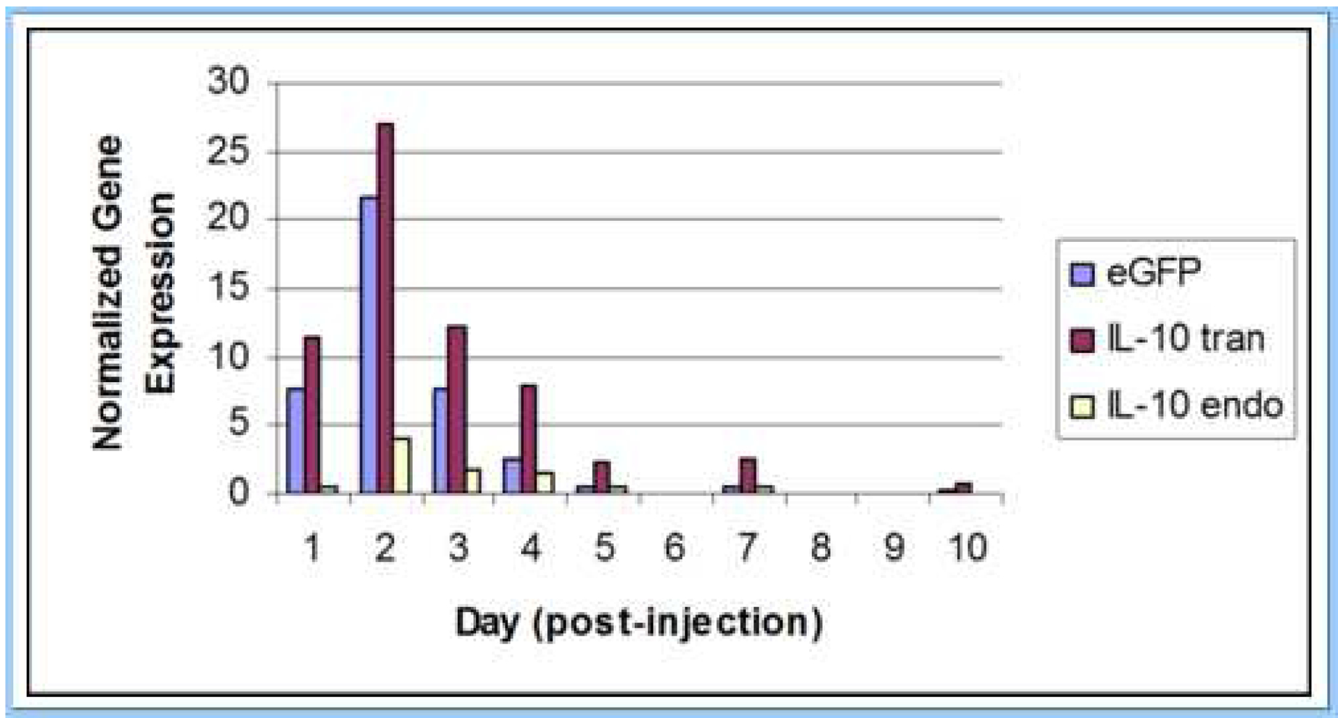
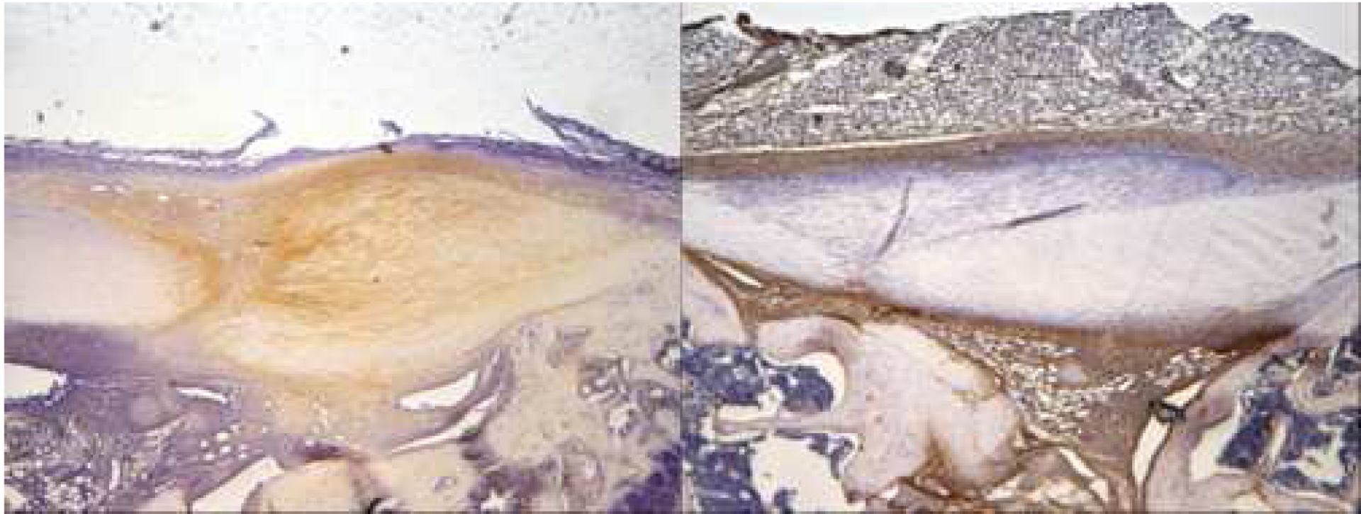
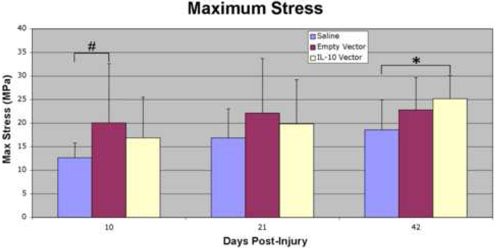
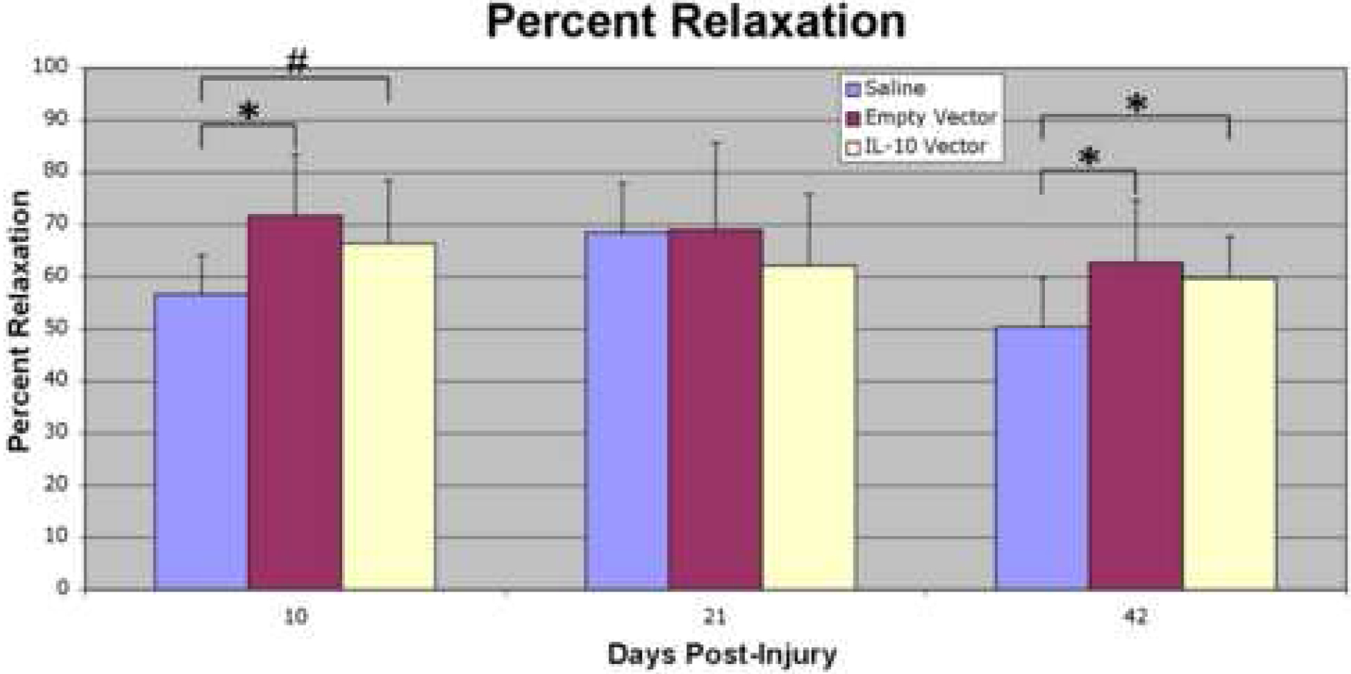
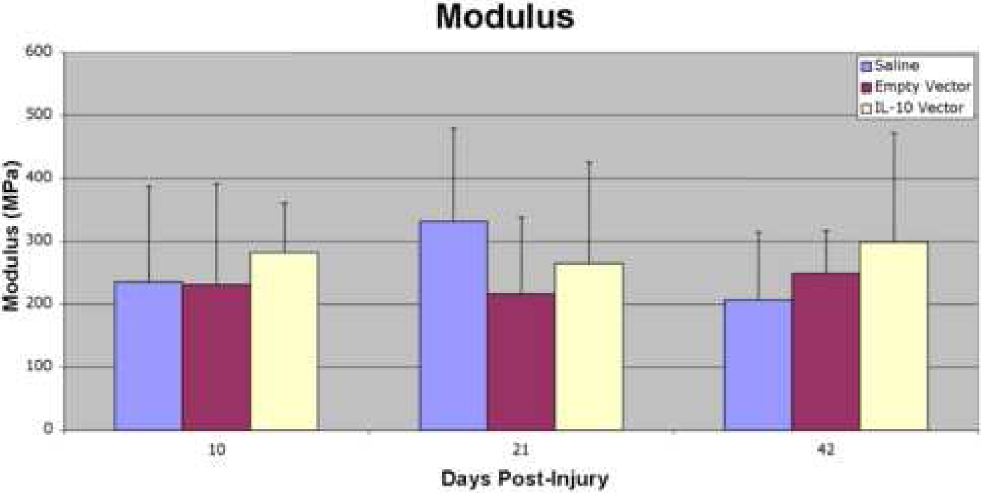
Methods: A murine model of patellar tendon injury was used and animals divided into 3 groups. Mice received bilateral patellar tendon injections with a lentiviral vector containing an IL-10 transgene (n = 34) or no transgene (n = 34). Control mice (n = 34) received injections of sterile saline. All animals then were subjected to bilateral, central patellar tendon injuries 2 days after injection and were killed at 5, 10, 21, and 42 days after injury. IL-10 content was analyzed by immunohistochemistry (n = 4/group). Tendon healing was evaluated by histology (n = 4/group) and biomechanical analysis (n = 10/group).
Results: Overexpression of IL-10 in patellar tendon was confirmed after injection of the lentiviral vector. IL-10 immunostaining was increased at day 10 in the IL-10 group relative to that in controls. Histologically, there was no significant difference in angular deviation between groups at day 21, but a trend toward decreased angular deviation in controls relative to that in empty vector group mice was seen at day 42. Biomechanically, the IL-10 group showed significantly increased maximum stress at day 42 relative to that in controls. Percent relaxation showed a trend toward an increase at day 10 and a significant increase at day 42 in the IL-10 group relative to that in controls.
Conclusions: This study demonstrates successful gene transfer of IL-10 into adult murine patellar tendon using a lentiviral vector. Although the effects of overexpression of IL-10 on adult tendon healing have not yet been fully elucidated, the current study may help to further clarify the mechanisms of tendon injury and repair.
Figures
Similar articles
-
Effect of a single versus serial platelet-rich plasma injection on the healing of acute patellar tendon defect: an experimental study.BMC Musculoskelet Disord. 2024 Aug 30;25(1):684. doi: 10.1186/s12891-024-07804-4. BMC Musculoskelet Disord. 2024. PMID: 39215319 Free PMC article.
-
Stem cells and bFGF in tendon healing: Effects of lentiviral gene transfer and long-term follow-up in a rat Achilles tendon defect model.BMC Musculoskelet Disord. 2016 Apr 5;17:148. doi: 10.1186/s12891-016-0999-6. BMC Musculoskelet Disord. 2016. PMID: 27048602 Free PMC article.
-
Use of Human Placenta-Derived Cells in a Preclinical Model of Tendon Injury.J Bone Joint Surg Am. 2019 Jul 3;101(13):e61. doi: 10.2106/JBJS.15.01381. J Bone Joint Surg Am. 2019. PMID: 31274724
-
Murine patellar tendon transplantation requires transosseous cerclage augmentation - development of a transplantation model for investigation of systemic and local drivers to healing.J Orthop Surg Res. 2019 Dec 2;14(1):410. doi: 10.1186/s13018-019-1475-4. J Orthop Surg Res. 2019. PMID: 31791383 Free PMC article.
-
CD44 deficiency improves healing tendon mechanics and increases matrix and cytokine expression in a mouse patellar tendon injury model.J Orthop Res. 2009 Oct;27(10):1386-91. doi: 10.1002/jor.20891. J Orthop Res. 2009. PMID: 19382192 Free PMC article.
Cited by
-
Therapeutic Efficacy of Intratendinous Delivery of Dexamethasone Using Porous Microspheres for Amelioration of Inflammation and Tendon Degeneration on Achilles Tendinitis in Rats.Biomed Res Int. 2020 Jan 21;2020:5052028. doi: 10.1155/2020/5052028. eCollection 2020. Biomed Res Int. 2020. PMID: 32090096 Free PMC article.
-
Preclinical assessment of IL-1β primed human umbilical cord mesenchymal stem cells for tendon functional repair through TGF-β/IL-10 signaling.Heliyon. 2023 Oct 21;9(11):e21411. doi: 10.1016/j.heliyon.2023.e21411. eCollection 2023 Nov. Heliyon. 2023. PMID: 37954299 Free PMC article.
-
Musculoskeletal regeneration and its implications for the treatment of tendinopathy.Int J Exp Pathol. 2013 Aug;94(4):293-303. doi: 10.1111/iep.12031. Epub 2013 Jun 17. Int J Exp Pathol. 2013. PMID: 23772908 Free PMC article. Review.
-
The roles of inflammatory mediators and immunocytes in tendinopathy.J Orthop Translat. 2018 Apr 14;14:23-33. doi: 10.1016/j.jot.2018.03.003. eCollection 2018 Jul. J Orthop Translat. 2018. PMID: 30035030 Free PMC article. Review.
-
Plasma Concentrations of Select Inflammatory Cytokines Predicts Pain Intensity 48 Hours Post-Shoulder Muscle Injury.Clin J Pain. 2020 Oct;36(10):775-781. doi: 10.1097/AJP.0000000000000861. Clin J Pain. 2020. PMID: 32675582 Free PMC article.
References
-
- Baratz ME, Schmidt CC, Hughes TB. Extensor Tendon Injuries. In: Green DP, Hotchkiss RN, Pederson WC, Wolfe SW, eds. Green’s Operative Hand Surgery. Vol. 1, 5th ed. Philadelphia: Elsevier Churchill Livingstone, 2005: 187–218.
-
- Boyer MI. Flexor Tendon Injury: Acute Injuries. In: Green DP, Hotchkiss RN, Pederson WC, Wolfe SW, eds. Green’s Operative Hand Surgery. Vol. 1, 5th ed. Philadelphia: Elsevier Churchill Livingstone, 2005: 219–240.
-
- Beredjiklian PK. Biologic aspects of flexor tendon laceration and repair. J Bone Joint Surg Am 2003; 85: 539–550. - PubMed
-
- Bruner S, Wittemann M, Jester A, Blumenthal K, Germann G. Dynamic splinting after extensor tendon repair in zones V to VII. J Hand Surg [Br] 2003; 28: 224–227. - PubMed
-
- Chow JA, Dovelle S, Thomes LJ, Ho PK, Saldana J. A comparison of results of extensor tendon repair followed by early controlled mobilisation versus static immobilisation. J Hand Surg [Br] 1989; 14: 18–20. - PubMed
Publication types
MeSH terms
Substances
Grants and funding
LinkOut - more resources
Full Text Sources
Medical
