. 2008 Dec 23;18(24):1917-21.
doi: 10.1016/j.cub.2008.10.029. Epub 2008 Dec 11.

ATP consumption by mammalian rod photoreceptors in darkness and in light

Affiliations

ATP consumption by mammalian rod photoreceptors in darkness and in light

Haruhisa Okawa et al. Curr Biol. .

Abstract

Why do vertebrates use rods and cones that hyperpolarize, when in insect eyes a single depolarizing photoreceptor can function at all light levels? We answer this question at least in part with a comprehensive assessment of ATP consumption for mammalian rods from voltages and currents and recently published physiological and biochemical data. In darkness, rods consume 10(8) ATP s(-1), about the same as Drosophila photoreceptors. Ion fluxes associated with phototransduction and synaptic transmission dominate; as in CNS, the contribution of enzymes of the second-messenger cascade is surprisingly small. Suppression of rod responses in daylight closes light-gated channels and reduces total energy consumption by >75%, but in Drosophila light opens channels and increases consumption 5-fold. Rods therefore provide an energy-efficient mechanism not present in rhabdomeric photoreceptors. Rods are metabolically less "costly" than cones, because cones do not saturate in bright light and use more ATP s(-1) for transducin activation and rhodopsin phosphorylation. This helps to explain why the vertebrate retina is duplex, and why some diurnal animals like primates have a small number of cones, concentrated in a region of high acuity.


Figures
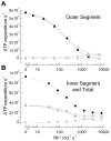
Figure 1
Figure 1
Principal contributors to ATP consumption in mammalian rod over the physiological range of steady light intensities. (A) Outer segment. ATP required for extrusion of Na+ entering cGMP-gated channels (■), transducin GTP hydrolysis and rhodopsin phosphorylation (□), synthesis of cGMP (○), and sum of all of these processes (X). (B) Inner segment and total rod ATP consumption. ATP required for extrusion of Na+ entering through ih channels (□), extrusion of Ca2+ entering voltage-gated channels at synaptic terminal (○), sum of ATP for Na+ and Ca2+ extrusion (△), and sum of ATP turnover in whole rod (●). For the global sum, we used light intensities at which voltage responses had been recorded (Fig. 2B) and estimated photocurrents by interpolation from step response-intensity data in Woodruff et al. [10]. See text and Supplemental Material for details of calculations.
Figure 2
Figure 2
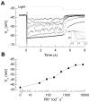
Perforated-patch current-clamp recordings of membrane potential from mouse rod photoreceptors in dark-adapted retinal slices. (A) Response waveforms to 5 s light steps of intensities 33, 83, 210, 520, 1300, 3200, and 8100 Rh* rod−1 s−1. Data from 8 cells were averaged individually for each background light intensity and were corrected for a measured liquid junction potential of ~10 mV. Average input resistance was 5 GΩ and average access resistance, 300 MΩ. Inset: Same responses at higher temporal resolution showing rapid relaxation or “nose” in voltage waveform at high light intensities caused by activation of ih. (B) Response-intensity curve of voltage response, averaged during the interval 4.5–5 s from the responses in A, as function of steady light intensity. Methods: Experiments were conducted in accordance with protocols approved by institutional IACUC committees. Light-evoked membrane potential changes during current-clamp recordings were measured in response to background light steps of 5 sec duration delivered from an LED (λmax ~ 470 nm). To estimate the number of rhodopsin molecules activated per flash, we measured the light intensity of a 520 μm spot focused on the slice preparation by the 20X 0.75NA (Nikon) condenser objective using a calibrated photodiode (United Detector Technologies, San Diego, CA). Light intensities were converted to equivalent 501 nm photons by convolving the power-scaled spectral output of the LED with the normalized spectral sensitivity curve for mouse rhodopsin. These were then converted to Rh* s−1 by estimating the collecting area of rod photoreceptors in the experimental setup. Dim flashes were delivered during suction electrode recordings from rod outer segments in clusters [42], and the mean Rh* per rod was determined from the scaling of the time-dependent variance to the mean response [43]. Based on these factors we estimated the rod collecting area in the experimental setup to be 0.18 μm2 (n = 6 rods).
Figure 3
Figure 3
Maximal and minimal ATP expenditure required for the biochemistry of the transduction cascade and by ion extrusion in a mouse rod in darkness and in bright illumination. The letters D and L indicate darkness and bright light. See text.

Similar articles

Cited by

References

    1. Hardie RC, Raghu P. Visual transduction in Drosophila. Nature. 2001;413:186–193. - PubMed
    1. Fain GL. Sensory Transduction. Sunderland, MA: Sinauer; 2003.
    1. Niven JE, Vahasoyrinki M, Juusola M. Shaker K(+)-channels are predicted to reduce the metabolic cost of neural information in Drosophila photoreceptors. Proc Biol Sci . 2003;270(Suppl 1):S58–61. - PMC - PubMed
    1. Attwell D, Laughlin SB. An energy budget for signaling in the grey matter of the brain. Journal of Cerebral Blood Flow and Metabolism. 2001;21 - PubMed
    1. Niven JE, Anderson JC, Laughlin SB. Fly photoreceptors demonstrate energy-information trade-offs in neural coding. PLoS Biology. 2007;5:828–840. (e116) - PMC - PubMed

Publication types

Substances