Evidence for a calcification process in the trabecular meshwork
- PMID: 19084518
- PMCID: PMC2670947
- DOI: 10.1016/j.exer.2008.11.027
Evidence for a calcification process in the trabecular meshwork
Abstract
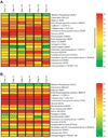
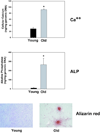
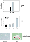
The human trabecular meshwork (TM) expresses many genes that have been associated with physiological (bone, cartilage, teeth) and pathological (vascular systems, kidney) calcification. In particular, the TM highly expresses the inhibitor of calcification Matrix Gla (MGP) gene, which encodes a vitamin K-dependent protein that requires post-translational activation to inhibit the formation of calcium precipitates. TM cells have high activity of the activating gamma-carboxylase enzyme and produce active MGP. Silencing MGP increases the activity of alkaline phosphatase (ALP), an enzyme of the matrix vesicles and marker of calcification. Overexpressing MGP reduces the ALP activity induced by bone morphogenetic 2 (BMP2), a potent inducer of calcification. In this review we gathered evidence for the existence of a mineralization process in the TM. We selected twenty regulatory calcification genes, reviewed their functions in their original tissues and looked at their relative abundance in the TM by heat maps derived from existing microarrays. Although results are not yet fully conclusive and more experiments are needed, examining TM expression in the light of the calcification literature brings up many similarities. One such parallel is the role of mechanical forces in bone induction and the high levels of mineralization inhibitors found in the constantly mechanically stressed TM. During the next few years, examination of other calcification-related regulatory genes and pathways, as well as morphological examination of knockout animals, would help to elucidate the relevance of a calcification process to TM's overall function.
Figures


References
-
- Addison WN, Azari F, Sorensen ES, Kaartinen MT, McKee MD. Pyrophosphate inhibits mineralization of osteoblast cultures by binding to mineral, up-regulating osteopontin, and inhibiting alkaline phosphatase activity. J. Biol. Chem. 2007;282:15872–15883. - PubMed
-
- Ameye L, Young MF. Mice deficient in small leucine-rich proteoglycans: novel in vivo models for osteoporosis, osteoarthritis, Ehlers-Danlos syndrome, muscular dystrophy, and corneal diseases. Glycobiology. 2002;12:107R–116R. - PubMed
-
- Anderson HC. Matrix vesicles and calcification. Curr. Rheumatol. Rep. 2003;5:222–226. - PubMed
-
- Ando Y, Ichihara N, Takeshita S, Saito Y, Kikuchi T, Wakasugi N. Histological and ultrastructural features in the early stage of Purkinje cell degeneration in the cerebellar calcification (CC) rat. Exp. Anim. 2004;53:81–88. - PubMed
Publication types
MeSH terms
Substances
Grants and funding
LinkOut - more resources
Full Text Sources
Medical
Miscellaneous

