Comparative Study
. 2008 Dec;5(12):1726-34.
doi: 10.1016/j.hrthm.2008.09.008. Epub 2008 Sep 6.

Role of late sodium current in modulating the proarrhythmic and antiarrhythmic effects of quinidine

Affiliations
Comparative Study

Role of late sodium current in modulating the proarrhythmic and antiarrhythmic effects of quinidine

Lin Wu et al. Heart Rhythm. 2008 Dec.

Abstract

Background: Quinidine is used to treat atrial fibrillation and ventricular arrhythmias. However, at low concentrations, it can induce torsade de pointes (TdP).

Objective: The purpose of this study was to examine the role of late sodium current (I(Na)) as a modulator of the arrhythmogenicity of quinidine in female rabbit isolated hearts and cardiomyocytes.

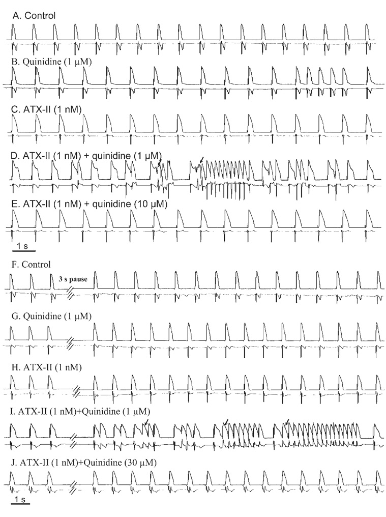
Methods: Epicardial and endocardial monophasic action potentials (MAPs), ECG signals, and ion channel currents were measured. The sea anemone toxin ATX-II was used to increase late I(Na).

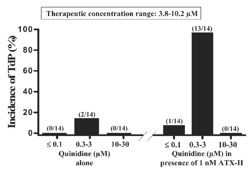
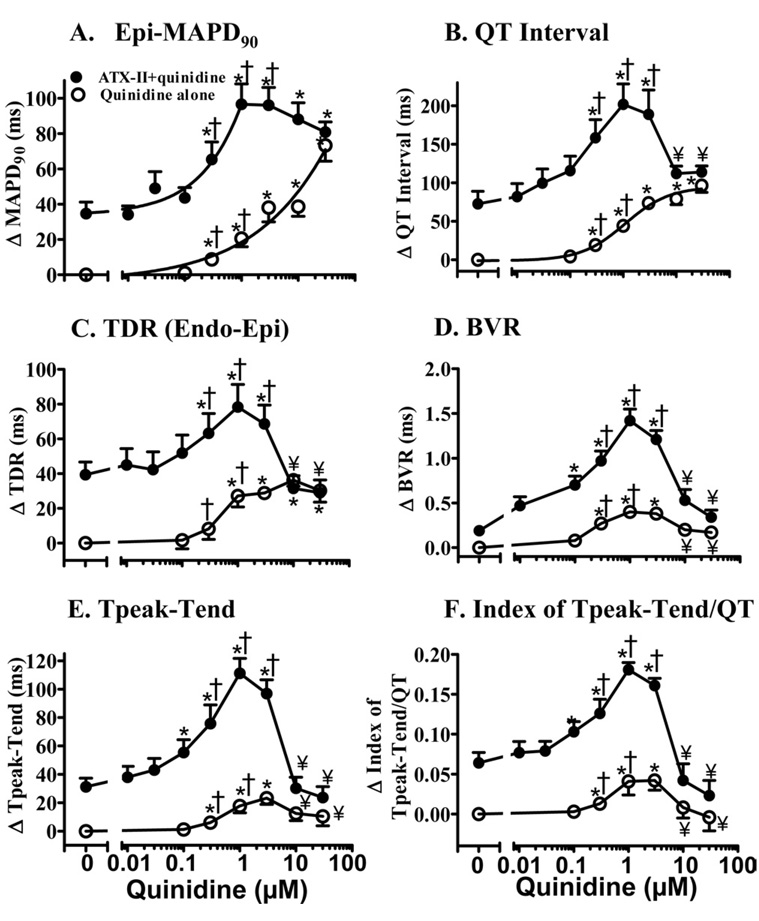
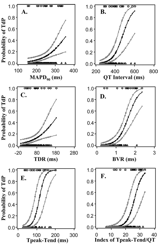
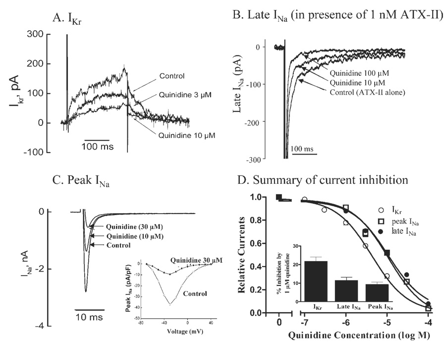
Results: Quinidine had concentration-dependent and often biphasic effects on measures of arrhythmogenicity. Quinidine increased the duration of epicardial MAP (MAPD(90)), QT interval, transmural dispersion of repolarization (TDR), and ventricular effective refractory period. Beat-to-beat variability of MAPD(90) (BVR), the interval from peak to end of the T wave (Tpeak-Tend) and index of Tpeak-Tend/QT interval were greater at 0.1 to 3 micromol/L than at 10-30 micromol/L quinidine. In the presence of 1 nmol/L ATX-II, quinidine caused significantly greater concentration-dependent and biphasic changes of Tpeak-Tend, TDR, BVR, and index of Tpeak-Tend/QT interval. Quinidine (1 micromol/L) induced TdP in 2 and 13 of 14 hearts in the absence and presence of ATX-II, respectively. Increases of BVR, index of Tpeak-Tend/QT interval, and Tpeak-Tend were associated with quinidine-induced TdP. Quinidine inhibited I(Kr), peak I(Na), and late I(Na) with IC(50)s of 4.5 +/- 0.3 micromol/L, 11.0 +/- 0.7 micromol/L, and 12.0 +/- 0.7 micromol/L.

Conclusion: Quinidine had biphasic proarrhythmic effects in the presence of ATX-II, suggesting that late I(Na) is a modulator of the arrhythmogenicity of quinidine. Enhancement of late I(Na) increased proarrhythmia caused by low but not high concentrations of quinidine.


Figures

Figure 1
Figure 1
Concentration-dependent biphasic proarrhythmic and antiarrhythmic effects of quinidine in causing torsade de pointes (TdP) in the absence (left) and presence (right) of 1 nmol/L ATX-II in female rabbit isolated hearts paced at 1 Hz. Numbers in parentheses indicate the number of hearts with TdP and the total number of hearts studied. Therapeutic concentrations range of quinidine is given in the box at the top of the figure.
Figure 2
Figure 2
Representative records of concentration-dependent biphasic proarrhythmic and antiarrhythmic effects of quinidine in the presence of 1 nmol/L ATX-II in a female rabbit heart paced at 1 Hz. Monophasic action potentials (top record in each panel) and ECG (bottom record in each panel) were simultaneously recorded. Drug effects recorded during constant pacing at 1 Hz (A–E) and after a 3-second pause (F–J) are shown. Arrowsindicate spontaneous or 3-second pause-triggered episodes of torsade de pointes.
Figure 3
Figure 3
Concentration–response relationships for quinidine in increasing epicardial MAPD90 (A), QT interval (B), transmural dispersion of MAPD90 (TDR, C), beat-to-beat variability of MAPD90 (BVR, D), Tpeak-Tend (E), and index of (Tpeak-Tend)/QT interval (F) in the absence (open symbols) and the presence (solid symbols) of 1 nmol/L ATX-II. Values are calculated as the changes from baseline in each heart and are presented as mean ± SEM. Baseline values (control) of these parameters are listed in Table 1 [available as online supplement]. *P <.05 quinidine alone vs control, or ATX-II + quinidine vs ATX-II alone. ¥P <.05 vs ATX-II + 1 µmol/L quinidine. †TdP occurred at concentrations indicated.
Figure 4
Figure 4
Relationships between an increase in the value of each of six electrophysiologic parameters and the probability of torsade de pointes (TdP) in a female rabbit isolated heart for all tested conditions (control, quinidine, ATX-II + quinidine). Symbols at the top of each plot indicate individual measured values of a given parameter in a heart with TdP (i.e., observed events). Symbols at the bottom of each figure indicate measured values of the same parameter in a heart without TdP (i.e., observed nonevents). Measured values were fitted using a linear mixed model with a logit link function to determine the predicted probability of TdP at any value of the parameter within the range of values recorded experimentally. The 95% confidence intervals for the predicted probabilities are shown. Abbreviations as in Figure 3.
Figure 5
Figure 5
Concentration-dependent inhibitions of IKr, late and peak INa by quinidine in female rabbit ventricular myocytes. A: Representative IKr currents from a single cell in the absence of drug (control) and during superfusion with 3 and 10 µmol/L quinidine. B: Representative recordings from a single cell of late INa in control (1 nmol/L ATX-II alone) and during superfusion with 10 and 100 µmol/L quinidine in the continued presence of 1 nmol/L ATX-II. C: Representative recordings of peak INa from a single cell in the absence (control) and the presence of 10 and 30 µmol/L quinidine. Insetshows current–voltage curves in the absence (open circles) and the presence (solid circles) of 30 µmol/L quinidine. D: Summarized concentration–response relationships for inhibitions of IKr, peak and late INa by quinidine (n = 4 for each).

Similar articles

Cited by

References

    1. Grace AA, Camm AJ. Quinidine. N Engl J Med. 1998;338:35–45. - PubMed
    1. Iost N, Virag L, Varro A, et al. Comparison of the effect of class IA antiarrhythmic drugs on transmembrane potassium currents in rabbit ventricular myocytes. J Cardiovasc Pharmacol Ther. 2003;8:31–41. - PubMed
    1. Kaufman ES. Quinidine in short QT syndrome: an old drug for a new disease. J Cardiovasc Electrophysiol. 2007;18:665–666. - PubMed
    1. Milberg P, Tegelkamp R, Osada N, et al. Reduction of dispersion of repolarization and prolongation of postrepolarization refractoriness explain the antiarrhythmic effects of quinidine in a model of short QT syndrome. J Cardiovasc Electrophysiol. 2007;18:658–664. - PubMed
    1. Marquez MF, Salica G, Hermosillo AG, et al. Ionic basis of pharmacological therapy in Brugada syndrome. J Cardiovasc Electrophysiol. 2007;18:234–240. - PubMed

Publication types

MeSH terms