Comparative Study
. 2008 Dec;5(12):1735-42.
doi: 10.1016/j.hrthm.2008.09.015. Epub 2008 Sep 16.

Atrial-selective effects of chronic amiodarone in the management of atrial fibrillation

Affiliations
Comparative Study

Atrial-selective effects of chronic amiodarone in the management of atrial fibrillation

Alexander Burashnikov et al. Heart Rhythm. 2008 Dec.

Abstract

Background: Although amiodarone is one of the most effective pharmacologic agents used in clinical management of atrial fibrillation (AF), little is known about its differential effects in atrial and ventricular myocardium.

Objectives: This study sought to compare the electrophysiological effects of chronic amiodarone in atria and ventricles.

Methods: We compared the electrophysiological characteristics of coronary-perfused atrial and ventricular wedge preparations isolated from untreated and chronic amiodarone-treated dogs (amiodarone, 40 mg/kg/day for 6 weeks, n = 12).

Results: Chronic amiodarone prolonged action potential duration (APD(90)) predominantly in atria compared to ventricles and prolonged the effective refractory period (ERP) more than APD(90) in both ventricular and atrial preparations (particularly in the latter) due to the development of postrepolarization refractoriness. Amiodarone reduced dispersion of APD(90) in both atria and ventricles. Although the maximum rate of increase of the action potential upstroke (V(max)) was significantly lower in both atria and ventricles of amiodarone-treated hearts versus untreated controls, the reduction of V(max) was much more pronounced in atria. Amiodarone prolonged P-wave duration more significantly than QRS duration, reflecting greater slowing of conduction in atria versus ventricles. These atrioventricular distinctions were significantly accentuated at faster activation rates. Persistent acetylcholine-mediated AF could be induced in only 1 of 6 atria from amiodarone-treated versus 10 of 10 untreated dogs.

Conclusion: Our results indicate that under the conditions studied, chronic amiodarone has potent atrial-predominant effects to depress sodium channel-mediated parameters and that this action of the drug is greatly potentiated by its ability to prolong APD predominantly in the atria, thus contributing to its effectiveness to suppress AF.


Figures

Figure 1
Figure 1
Chronic amiodarone prolongs APD90 predominantly in atria vs. ventricles. Upper panel: Superimposed action potential recorded from pectinate muscle in atrial and ventricular epicardial preparations isolated from untreated control (C) and amiodarone-treated dogs. Dashed lines denote the level of APD90 and the numbers indicate the changes in APD90 induced by chronic amiodarone. Lower panel: Graphs plot the average APD90 data. CT – crista terminalis, PM – pectinate muscle, M cell – M cell region, Epi – epicardium. * p<0.05 - vs. respective controls. N=7-16. CL = 500 ms.
Figure 2
Figure 2
Chronic amiodarone predominantly prolongs ERP in atria vs. ventricle due to predominant prolongation of atrial APD and the development of PRR primarily in atria. PRR is defined as the difference of APD75 and ERP in atria and APD90 and ERP in ventricles (ERP corresponds to APD75 in atria and APD90 in ventricles, contributing to a shorter ERP in atria). Upper panel: schematic illustration of the effects of chronic amiodarone to prolong ERP in atria and ventricles, through Na+ channel blockade. The arrows denote the position on the action potential corresponding to the end of the ERP in atria and ventricles and the effect of chronic amiodarone (Na+ channel block) to shift the end of the ERP predominantly in atria. C – Control. Bottom panel: Summary data from pectinate muscles in atria and epicardial region in ventricles at a CL of 500 ms (n = 3-10). * p<0.05 - vs. respective controls; † p<0.05 vs. APD75. See “Discussion” for details.
Figure 3
Figure 3
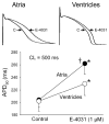
Why does amiodarone cause a preferential prolongation of atrial APD? The selective IKr blocker E-4031 (1 μM) prolongs APD90 in atria to a much greater extent than in ventricular preparations isolated from untreated control dogs. Atria – pectinate muscle; Ventricles – M cell region. * p < 0.05 vs. respective untreated control. † p<0.05 vs. respective ventricles. n=5-7.
Figure 4
Figure 4. Chronic amiodarone produces a rate-dependent depression of maximum rate of rise of the action potential upstroke (Vmax) predominantly in atria vs. ventricles
A: Atrial-predominant rate-dependent depression of Vmax following chronic amiodarone. CL = cycle length. B: Plot of normalized reduction in Vmax (n= 4-14). “Atria” represent combined PM and CT data. “Ventricles” represent combined epicardial and M cell data. * p < 0.05 vs. untreated control. † p<0.001 vs. control. C: Mechanism contributing to atrial selectivity of chronic amiodarone to depress Vmax at rapid activation rates: Greater APD prolongation resulting in greater reduction or elimination of diastolic interval (during which recovery from Na+ channel block primarily occurs) and a more positive take off potential. Dashed line indicates RMP in atria.
Figure 5
Figure 5
Chronic amiodarone prolongs the duration of “P wave” complex in atrial preparations to a greater extent than the duration of “QRS” complex in ventricular preparations, reflecting a greater and rate-dependent slowing of conduction velocity in atria vs. ventricles. CL – cycle length. * p < 0.05 vs. control. † p<0.05 vs respective CL = 500 ms. n=6-10.
Figure 6
Figure 6. Chronic amiodarone prevents induction of AF in the presence of acetylcholine (ACh) by prolonging APD and reducing excitability, preventing rapid 1:1 activation
Upper panel: ECG and action potential (AP) tracings during pacing at CL of 100 and 70 ms in the presence of ACh leads to AF induction in an atrium isolated from an untreated dog. Bottom panel: ECG and AP tracings during acceleration of pacing rate, leading to activation failure at a CL of 150 ms without arrhythmia induction in an atrial preparation isolated from a chronic amiodarone-treated dog.

Comment in

References

    1. Nattel S, Carlsson L. Innovative approaches to anti-arrhythmic drug therapy. Nat Rev Drug Discov. 2006;5:1034–49. - PubMed
    1. Burashnikov A, Di Diego JM, Zygmunt AC, et al. Atrium-selective sodium channel block as a strategy for suppression of atrial fibrillation: differences in sodium channel inactivation between atria and ventricles and the role of ranolazine. Circulation. 2007;116:1449–57. - PMC - PubMed
    1. Singh BN. Amiodarone: a multifaceted antiarrhythmic drug. Curr Cardiol Rep. 2006;8:349–55. - PubMed
    1. Kodama I, Kamiya K, Toyama J. Amiodarone: ionic and cellular mechanisms of action of the most promising class III agent. Am J Cardiol. 1999;84:20R–8R. - PubMed
    1. Goldschlager N, Epstein AE, Naccarelli GV, et al. A practical guide for clinicians who treat patients with amiodarone: 2007. Heart Rhythm. 2007;4:1250–9. - PubMed

Publication types

MeSH terms