

Phenotype-genotype correlations in autosomal dominant retinitis pigmentosa caused by RHO, D190N
- PMID: 19085385
- PMCID: PMC2749948
- DOI: 10.1080/02713680802484645
Phenotype-genotype correlations in autosomal dominant retinitis pigmentosa caused by RHO, D190N
Abstract
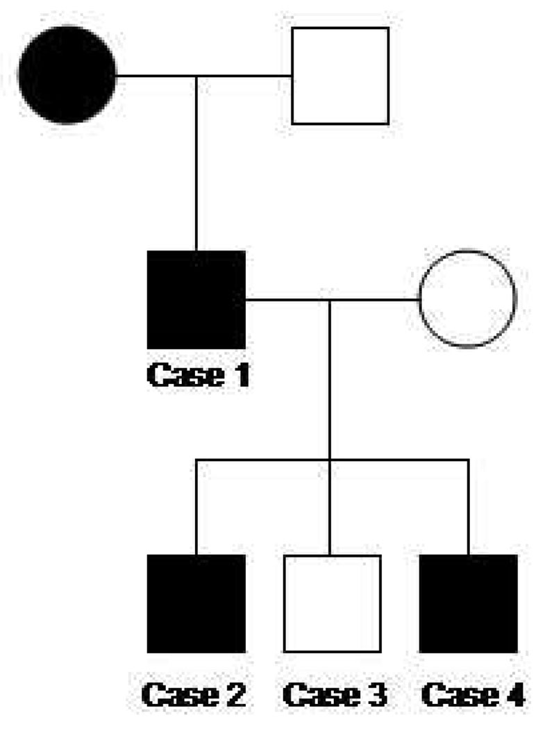
Purpose: To phenotype a family with RHO (Asp190Asn or D190N) dominantly inherited retinitis pigmentosa (RP) and to describe an approach to surveying affected families.
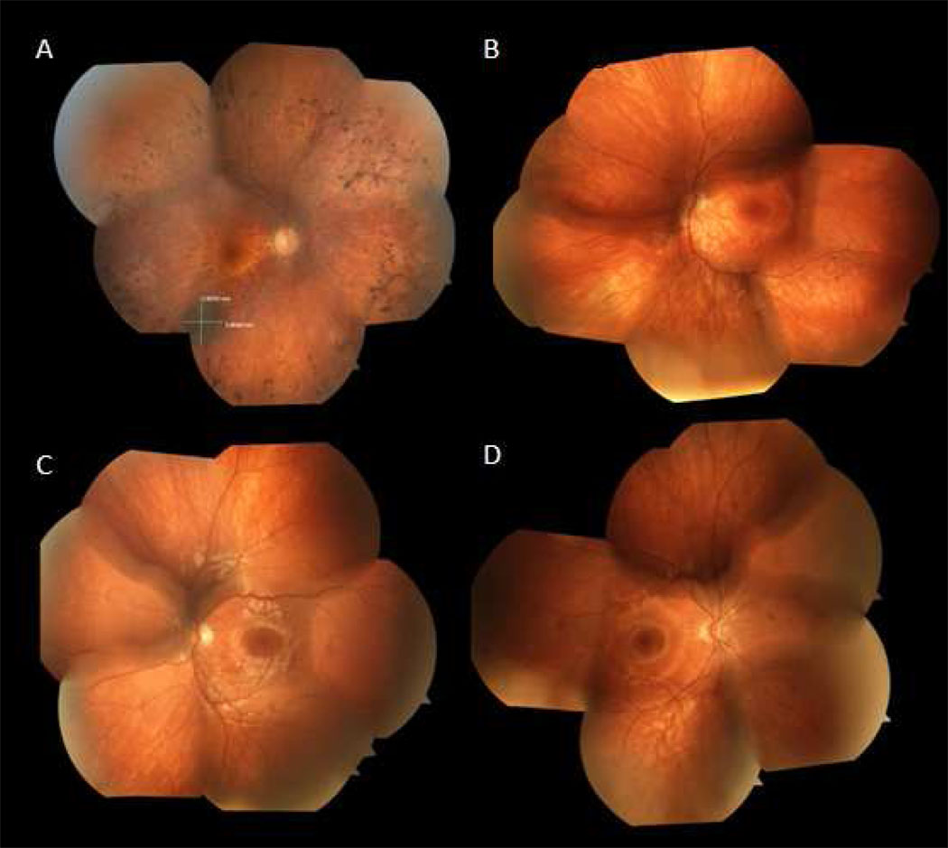
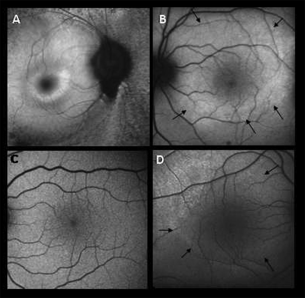
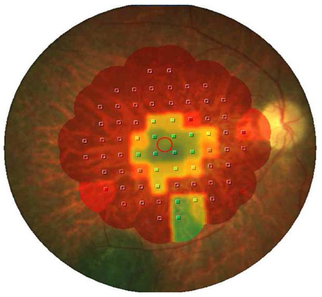
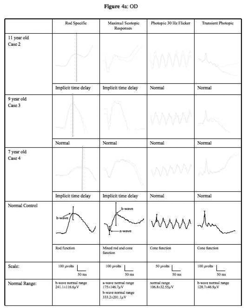
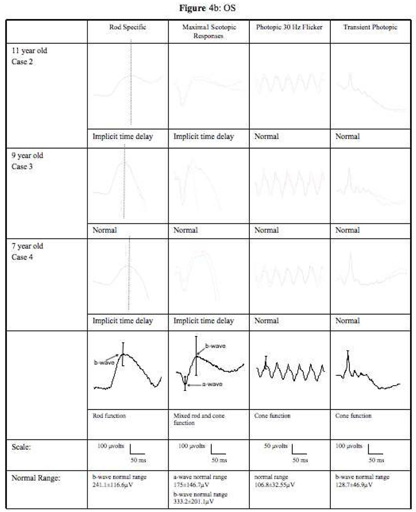
Methods: Four patients from a family with a history of autosomal dominant RP had complete clinical examinations and underwent full-field electroretinography (ERG), fundus autofluorescence (AF) imaging, and genetic testing. One patient had microperimetry (MP) mapping.
Results: The patients' ages ranged from 6 years to 47 years. The proband, the father, had fundoscopic findings typical of RP. A small hyperfluorescent ring centered at the fovea was apparent on AF. MP showed preservation of central 7 degrees of visual field within this ring. The three children were all asymptomatic with visual acuity of 20/15 in each eye. One child had mild retinal pigment epithelium migration on fundoscopy; the other two children had normal fundoscopic examinations. Two children showed increased parafoveal AF. In the two affected children, average ERG b-wave implicit times were delayed in scotopic conditions, and maximal ERG tracings had abnormal waveforms. Genetic analysis confirmed that two of three asymptomatic children carried the D190N allele.
Conclusions: Patients with RHO (D190N) autosomal dominant retinitis pigmentosa (adRP) can show classic signs of RP on fundus examination and may be able to maintain good central visual acuity into adulthood. By combining clinical examination with AF imaging and electrophysiology, it is possible to offer presymptomatic clinical evaluation to families with this RP.
Figures
References
-
- Berson EL. Retinitis Pigmentosa: The Friedenwald Lecture. Invest Opthal Vis Sci. 1993;34:1655–1676. - PubMed
-
- Bunker CH, Berson EL, Bromley WC, Hayes RP, Roderick TH. Prevalence of retinitis pigmentosa in Maine. American J of Opthalmol. 1984;97:357–365. - PubMed
-
- Tsang SH, Gouras P. Chapter 2. Molecular Physiology and Pathology of the Retina. In: Tasman W, Jaeger EA, editors. Duane's Clinical Opthalmology. Philadelphia: J.B. Lippincott; 1996.
Publication types
MeSH terms
Substances
Grants and funding
LinkOut - more resources
Full Text Sources
Research Materials