. 2009 Mar;30(3):379-90.
doi: 10.1002/humu.20868.

Classic and atypical fibrodysplasia ossificans progressiva (FOP) phenotypes are caused by mutations in the bone morphogenetic protein (BMP) type I receptor ACVR1

Affiliations

Classic and atypical fibrodysplasia ossificans progressiva (FOP) phenotypes are caused by mutations in the bone morphogenetic protein (BMP) type I receptor ACVR1

Frederick S Kaplan et al. Hum Mutat. 2009 Mar.

Abstract

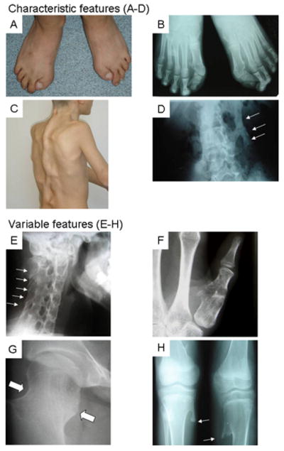
Fibrodysplasia ossificans progressiva (FOP) is an autosomal dominant human disorder of bone formation that causes developmental skeletal defects and extensive debilitating bone formation within soft connective tissues (heterotopic ossification) during childhood. All patients with classic clinical features of FOP (great toe malformations and progressive heterotopic ossification) have previously been found to carry the same heterozygous mutation (c.617G>A; p.R206H) in the glycine and serine residue (GS) activation domain of activin A type I receptor/activin-like kinase 2 (ACVR1/ALK2), a bone morphogenetic protein (BMP) type I receptor. Among patients with FOP-like heterotopic ossification and/or toe malformations, we identified patients with clinical features unusual for FOP. These atypical FOP patients form two classes: FOP-plus (classic defining features of FOP plus one or more atypical features) and FOP variants (major variations in one or both of the two classic defining features of FOP). All patients examined have heterozygous ACVR1 missense mutations in conserved amino acids. While the recurrent c.617G>A; p.R206H mutation was found in all cases of classic FOP and most cases of FOP-plus, novel ACVR1 mutations occur in the FOP variants and two cases of FOP-plus. Protein structure homology modeling predicts that each of the amino acid substitutions activates the ACVR1 protein to enhance receptor signaling. We observed genotype-phenotype correlation between some ACVR1 mutations and the age of onset of heterotopic ossification or on embryonic skeletal development.


Figures

Figure 1
Figure 1. Characteristic and Variable Features of Classic FOP
Composite of characteristic (A-D) and common variable (E-H) features of classic FOP. A photograph (A) and radiograph (B) of the feet in a classically-affected 15 year-old boy shows short, malformed monophalangic great toes. A photograph of his back (C) and a radiograph of his lumbar spine (D) reveal ribbons, sheets, and plates of heterotopic bone (D, arrows). Lower panel collage (E-H) from several affected individuals depicts common variable features of classic FOP including orthotopic fusion of sub-axial facet joints of the cervical spine (E, arrows) prior to the onset of heterotopic ossification, short monophalangic malformed thumb (F), short broad femoral neck (G, arrows), and proximal medial tibial osteochondromas (H, arrows).
Figure 2
Figure 2. Digital Malformations in FOP Variants
All FOP variants patients whose images are depicted here (A-H) had heterozygous missense mutations at either codon 328 (A-E) or codon 536 (F-H) in the kinase domain of ACVR1 in contrast to all classically-affected individuals with FOP who had a recurrent mutation in ACVR1 at codon 206 (R206H) (Figure 1); protein RefSeq NP_001096.1. All three variants with the G328R mutation (A, B, plus members of family #9, not shown) had either normal great toes or minimal malformations, while the hands were normal. All four variants with either the G328W mutation (C; plus patient #12, not shown) or the G328E mutation (D, E) had severe truncation deformities of multiple digits (C-E) and/or syndactyly (C, D). Patients #13 and #14 (D, E; G328E mutation) had slightly different malformation patterns from each other: patient #13 (D) had severe truncation of the great toes, whereas patient #14 (E) had more severe reduction deficits of the posterior digits. The hand malformations were similar in each, although patient #14 (E) was missing a post-axial digit on both hands. Both patients lacked nails in all severely affected digits. Three variants with the G356D mutation (F, G, H) had severe truncation malformations of the thumbs and great toes, although variable degrees of terminal symphalangism were noted and the digital truncations of patient #16 (F) were asymmetric in the hands and feet.
Figure 3
Figure 3. Position and Conservation of ACVR1 Amino Acid Changes
ACVR1 encodes a 509 amino acid protein that contains a ligand binding region, a transmembrane (TM) domain, a glycine-serine rich region (GS), and a protein kinase domain. The numbers below the protein representation indicate the amino acid codons included in each identified domain; the protein initiation codon is codon 1 (RefSeq NP_001096.1). The relative positions of all identified mutations are shown with the altered amino acids in bold with light shading. Each mutation in the ACVR1 gene occurs in an identical amino acid at the corresponding position of ACVR1/ALK2 across species (darker shading). Clustal W was used for multiple protein sequence alignment. The schematics are drawn approximately to scale.
Figure 4
Figure 4. Sites of FOP mutations in a structured-based homology model of the ACVR1 receptor kinase domain
Panels show models of the wild type ACVR1 protein with specific amino acids that are implicated in structural changes as a result of mutations are indicated. The protein initiation codon is codon 1 (RefSeq NP_001096.1). A. Mutation sites within the GS regulatory region. Arginine 206, which forms an ion pair with aspartate 269 (dashed lines), is substituted with histidine in all patients with classic FOP and six of eight patients (#1-6) with FOP-plus. The adjacent residue, glutamine 207, is substituted with glutamate in FOP-plus patient #7. A three-nucleotide deletion replaces proline 197 and phenylalanine 198 with a leucine residue in variant FOP patient #20. The surface of the FKBP12 binding protein at the binding protein-receptor interface is depicted in grey. B, C. Multiple Glycine 328 missense mutations. Seven FOP variants (#8-14) had substitutions of glycine 328. Codon 328, in the protein kinase domain, resides in a loop at the bottom of a surface cavity bordering the GS loop and the N-terminal end of the αC helix. The surface of the kinase domain is depicted in green. For clarity, FKBP12 is not shown. The view in C is similar to panel B, with the FKBP12 binding site and GS loop rolled slightly forward toward the viewer. D. Mutation sites within the receptor kinase active site. Glycine 356 is substituted with aspartate in patients #15-18 (one with FOP-plus and three FOP variants) and arginine 375 with proline in FOP variant patient #19. The ion pair between arginine 375 and aspartate 354 blocks a cation binding site required for ATP hydrolysis by the enzyme. The lysine 235-glutamate 248 ion pair is conserved in all protein kinases and modulates enzyme activity by altering active site conformation.

References

    1. Ahn J, de la Pena LS, Shore EM, Kaplan FS. Paresis of a bone morphogenetic protein-antagonist response in a genetic disorder of heterotopic skeletogenesis. J Bone Joint Surg (Am) 2003;85A:667–674. - PubMed
    1. Andreev K, Zenkel M, Kruse F, Junemann A, Schlotzer-Schrehardt U. Expression of bone morphogenetic proteins (BMPs), their receptors, and activins in normal and scarred conjunctiva: role of BMP-6 and activin-A in conjunctival scarring? Exp Eye Res. 2006;83:1162–1170. - PubMed
    1. Angley C, Kumar M, Dinsio KJ, Hall AK, Siegel RE. Signaling by bone morphogenetic proteins and Smad1 modulates the postnatal differentiation of cerebellar cells. J Neurosci. 2003;23:260–268. - PMC - PubMed
    1. Bakrania P, Efthymiou M, Klein JC, Salt A, Bunyan DJ, Wyatt A, Ponting CP, Mmartin A, Williams S, Lindley V, Gilmore J, Restori M, Robson AG, Neveu MM, Holder GE, Collin JRO, Robinson DO, Fardon P, Johansen-Berg H, Gerrelli D, Ragge NK. Mutations in BMP4 cause eye, brain, and digit developmental anomalies: Overlap between the BMP4 and Hedgehog signaling pathways. Am J Hum Genet. 2008;82:304–319. - PMC - PubMed
    1. Baur ST, Mai JJ, Dymecki SM. Combinatorial signaling through BMP receptor IB and GDF5: shaping of the distal mouse limb and the genetics of distal limb diversity. Development. 2000;127:605–619. - PubMed

Publication types

Substances