. 2009 Mar;49(3):960-8.
doi: 10.1002/hep.22697.

Functional linkage of cirrhosis-predictive single nucleotide polymorphisms of Toll-like receptor 4 to hepatic stellate cell responses

Affiliations

Functional linkage of cirrhosis-predictive single nucleotide polymorphisms of Toll-like receptor 4 to hepatic stellate cell responses

Jinsheng Guo et al. Hepatology. 2009 Mar.

Abstract

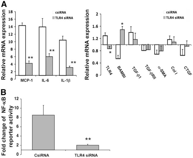
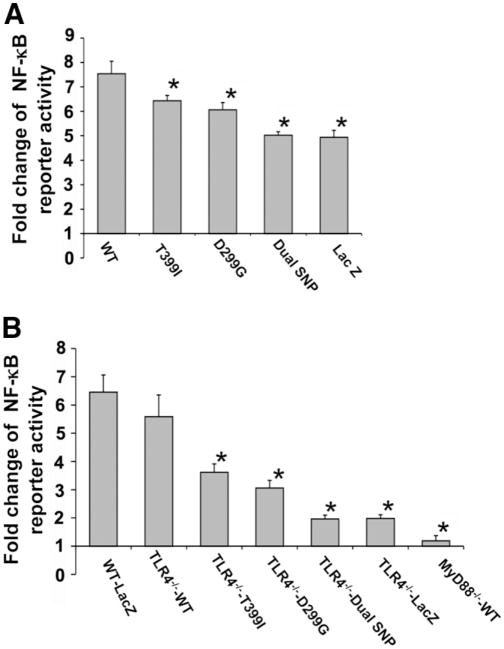
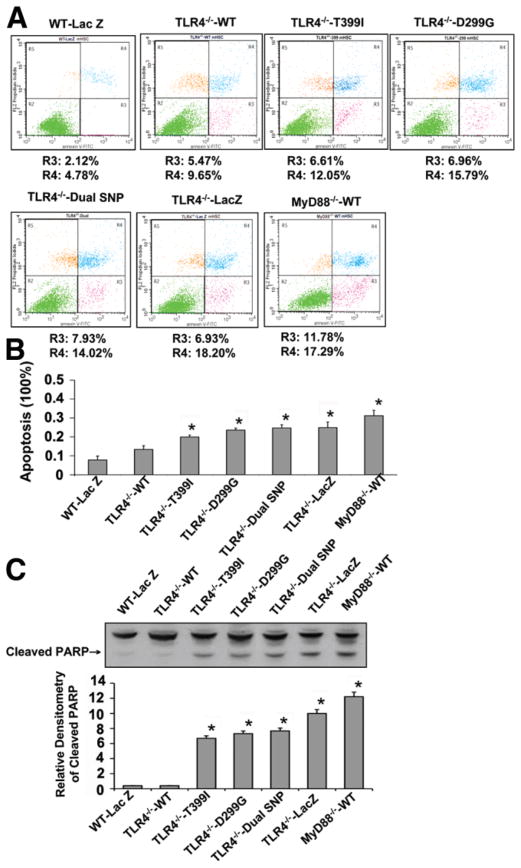
In a recent study, a single nucleotide polymorphism (SNP) of the Toll-like receptor 4 (TLR4) gene (c.1196C>T [rs4986791, p.T399I]) emerged as conferring protection from fibrosis progression compared to a major, wild-type (WT) CC allele (p.T399). The present study examined the functional linkage of this SNP, along with another common, highly cosegregated TLR4 SNP (c.896A>G [rs4986790, p.D299G]), to hepatic stellate cell (HSC) responses. Both HSCs from TLR4(-/-) mice and a human HSC line (LX-2) reconstituted with either TLR4 D299G and/or T399I complementary DNAs were hyporesponsive to lipopolysaccharide (LPS) stimulation compared to those expressing WT TLR4, as assessed by the expression and secretion of LPS-induced inflammatory and chemotactic cytokines (i.e., monocyte chemoattractant protein-1, interleukin-6), down-regulation of bone morphogenic protein and the activin membrane-bound inhibitor expression (an inhibitory transforming growth factor beta pseudoreceptor), and activation of a nuclear factor kappaB (NF-kappaB)-responsive luciferase reporter. In addition, spontaneous apoptosis, as well as apoptosis induced by pathway inhibitors of NF-kappaB, extracellular signal-regulated kinase (ERK), and phosphatidylinositol 3-kinase were greatly increased in HSCs from either TLR4(-/-) or myeloid differentiation factor 88(-/-) (a TLR adaptor protein) mice, as well as in murine HSCs expressing D299G and/or T399I SNPs; increased apoptosis in these lines was accompanied by decreased phospho-ERK and Bcl-2.

Conclusion: TLR4 D299G and T399I SNPs that are associated with protection from hepatic fibrosis reduce TLR4-mediated inflammatory and fibrogenic signaling and lower the apoptotic threshold of activated HSCs. These findings provide a mechanistic link that explains how specific TLR4 SNPs may regulate the risk of fibrosis progression.


Conflict of interest statement

Potential conflict of interest: Nothing to report.

Figures

Fig. 1
Fig. 1
TLR4 siRNA abrogates LPS responsiveness in LX-2 cells. Following transfection with TLR4 siRNAs alone for mRNA analysis by real-time PCR or cotransfection with an NF-κB responsive reporter plasmid for the assessment of NF-κB activation, LX-2 cells were stimulated with LPS (100 ng/mL) for 12 hours. (A) Relative mRNA expression of inflammatory cytokines and fibrogenic genes in LX-2 cells transfected with TLR4 siRNAs (TLR4 siRNA) or control siRNA (csiRNA) in response to LPS. Data are depicted relative to expression in cells without LPS or siRNA, which are assigned a value of 1. (B) Fold change of NF-κB responsive reporter activity in LX-2 cells transfected with TLR4 siRNAs or control siRNA in response to LPS. Data are depicted relative to expression in cells without LPS or siRNA, which are assigned a value of 1. TLR4 siRNA abrogated LPS-induced up-regulation of MCP-1 and IL-6, and down-regulation of BAMBI, coincident with a reduced responsiveness of NF-κB activity. Each column represents the mean ± SEM of n = 6 per group in three independent experiments. *P < 0.05; **P < 0.01 when compared with the control siRNA transfected cells.
Fig. 2
Fig. 2
TLR4 SNPs confer decreased NF-κB activation to LPS stimulation in HSCs. An NF-κB responsive reporter plasmid was cotransfected with TLR4 cDNAs into LX-2 cells or transfected into mHSCs stably reconstituted with human TLR4 cDNAs. The cells were stimulated with LPS (100 ng/mL) for 12 hours. The activation of NF-κB responsive reporter was determined with a dual-luciferase reporter assay system. (A) Fold change of NF-κB reporter activity in LX-2 cells transfected with TLR4 WT or SNP sequences, or a LacZ control vector. (B) Fold change of NF-κB reporter activity in TLR4−/− mouse HSCs transfected with TLR4 WT or SNP sequences, or a LacZ control vector, and in MyD88−/− HSCs reconstituted with WT human TLR4 sequence. Cells expressing either the single or dual SNPs had less NF-κB activation. Each column represents the mean ± SEM of n = 6 per group in more than three independent experiments. *P < 0.05 when compared with the WT TLR4 cDNA-transfected stellate cells.
Fig. 3
Fig. 3
TLR4 SNPs attenuate the response of NF-κB target genes in HSCs following LPS stimulation. LX-2 cells or TLR4−/− mHSC lines that were transiently or stably reconstituted with human TLR4 cDNAs were stimulated with 100 ng/mL LPS for 12 hours. The mRNA expression of inflammatory cytokines MCP-1 and IL-6, and BAMBI in LX-2 cells (A) and mHSCs (B) was determined by real-time quantitative PCR. The parallel changes in protein secretion of MCP-1 and IL-6 in the cell culture supernatants of LX-2 cells (C) and mouse HSCs (D) were assessed by ELISA. The fold changes of MCP-1 and IL-6 mRNAs after LPS exposure were reduced in the presence of single or dual SNPs, whereas the extent of BAMBI down-regulation was attenuated. Changes of α-smooth muscle actin and collagen type I mRNAs were not statistically different following reconstitution with TLR4 SNPs (not shown). Each column represents the mean ± SEM of n = 6 per group in three independent experiments. *P < 0.05 when compared with the WT TLR4 cDNA-transfected stellate cells.
Fig. 4
Fig. 4
TLR4 SNPs reduce mHSC growth and enhance growth inhibition by pathways inhibitors. TLR4−/− mHSC lines that were stably reconstituted with human TLR4 cDNAs were treated with PI3K inhibitor (LY 294002), ERK inhibitor (PD 98059), or NF-κB inhibitor (20 μM, 10 μM, and 5 μM, respectively). DNA synthesis was assessed by 3[H]-incorporation. (A) DNA synthesis in TLR4−/− mHSCs transfected with human TLR4 WT or SNP cDNAs and MyD88−/− HSCs reconstituted with human TLR4 WT. (B) Growth reduction rate following addition of a PI3K inhibitor, ERK inhibitor, or NF-κB inhibitor. Cells with either single or dual SNPs had slower cell growth and more growth reduction following pathway inhibition than WT TLR4 cells. Each column represents the mean ± SEM of n = 6 per group in three independent experiments. *P < 0.05 when compared with the WT TLR4 cDNA-transfected HSCs.
Fig. 5
Fig. 5
TLR4 SNPs increase the spontaneous apoptosis of mHSCs. Spontaneous cell apoptosis was assessed in TLR4−/− mHSC lines that were stably reconstituted with human TLR4 cDNAs by FACS and by western blot analysis of cleaved PARP. (A) FACS analysis of spontaneous apoptosis of TLR4−/− mouse HSCs transfected with human TLR4 cDNAs and MyD88−/− HSCs reconstituted with WT TLR4. (B) Apoptosis rate (including both the early apoptotic rate [R3] and late apoptotic rate [R4]) was determined by FACS analysis of each cell line. (C) Western blot analysis of cleaved PARP in each cell line (representative western blotted membrane and relative densitometric quantitation are shown). Each column represents the mean ± SEM of n = 3 per group in three independent experiments. *P < 0.01 when compared with WT TLR4 cDNA-transfected stellate cells. Cells expressing single or dual SNPs showed higher spontaneous apoptosis than WT TLR4-transfected cells.
Fig. 6
Fig. 6
TLR4 SNPs lower the apoptotic threshold of activated HSCs. TLR4−/− mHSC lines that were stably reconstituted with TLR4 cDNAs were either serum-starved for 24 hours or incubated with pathway inhibitors including PI3K inhibitor (LY 294002), ERK inhibitor (PD 98059), or NF-κB inhibitor for 12 hours (20 μM, 10 μM, and 5 μM, respectively). The apoptosis rate was analyzed by FACS. Cells expressing either single or dual TLR4 SNPs have higher apoptotic induction rates under serum starvation or pathway inhibition than WT TLR4-expressing cells. Each column represents the mean ± SEM of n = 3 per group in three independent experiments. *P < 0.01 when compared with WT TLR4 cDNA-transfected HSCs.
Fig. 7
Fig. 7
TLR4 SNPs alter the level of apoptosis-related proteins in mHSCs. TLR4−/− mHSC lines that were stably reconstituted with human TLR4 cDNAs were stimulated with 100 ng/mL LPS for 12 hours. Western blot of apoptosis-regulating proteins was performed, including total ERK (t-ERK), p-ERK, total Akt (t-Akt), p-Akt, Bcl-2, and Bax (representative western blotted membrane and relative densitometric quantitation to β-actin are shown). Two key downstream effectors regulating apoptosis (p-ERK and Bcl-2) were reduced in TLR4 knockout and T399I, D299G, dual D299G/T399I SNP-expressing stellate cells. The figure is a representative of three independent experiments. *P < 0.01 compared with WT TLR4 cDNA-transfected HSC. #P < 0.05 compared with the cells without LPS treatment.

References

    1. Poynard T, Bedossa P, Opolon P. Natural history of liver fibrosis progression in patients with chronic hepatitis C. The OBSVIRC, METAVIR, CLINIVIR, and DOSVIRC groups. Lancet. 1997;349:825–832. - PubMed
    1. Asselah T, Bieche I, Paradis V, Bedossa P, Vidaud M, Marcellin P. Genetics, genomics, and proteomics: implications for the diagnosis and the treatment of chronic hepatitis C. Semin Liver Dis. 2007;27:13–27. - PubMed
    1. Huang H, Shiffman ML, Friedman S, Venkatesh R, Bzowej N, Abar OT, et al. A 7 gene signature identifies the risk of developing cirrhosis in patients with chronic hepatitis C. Hepatology. 2007;46:297–306. - PubMed
    1. Arbour NC, Lorenz E, Schutte BC, Zabner J, Kline JN, Jones M, et al. TLR4 mutations are associated with endotoxin hyporesponsiveness in humans. Nat Genet. 2000;25:187–191. - PubMed
    1. Lorenz E, Mira JP, Frees KL, Schwartz DA. Relevance of mutations in the TLR4 receptor in patients with gram-negative septic shock. Arch Intern Med. 2002;162:1028–1032. - PubMed

Publication types

MeSH terms