. 2008 Dec:Chapter 19:Unit 19.9.
doi: 10.1002/0471143030.cb1909s41.

Three-dimensional tissue models of normal and diseased skin

Affiliations

Three-dimensional tissue models of normal and diseased skin

Mark W Carlson et al. Curr Protoc Cell Biol. 2008 Dec.

Abstract

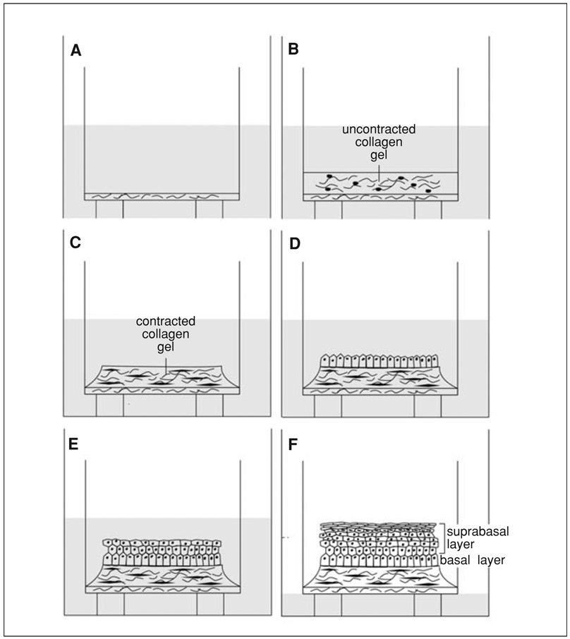
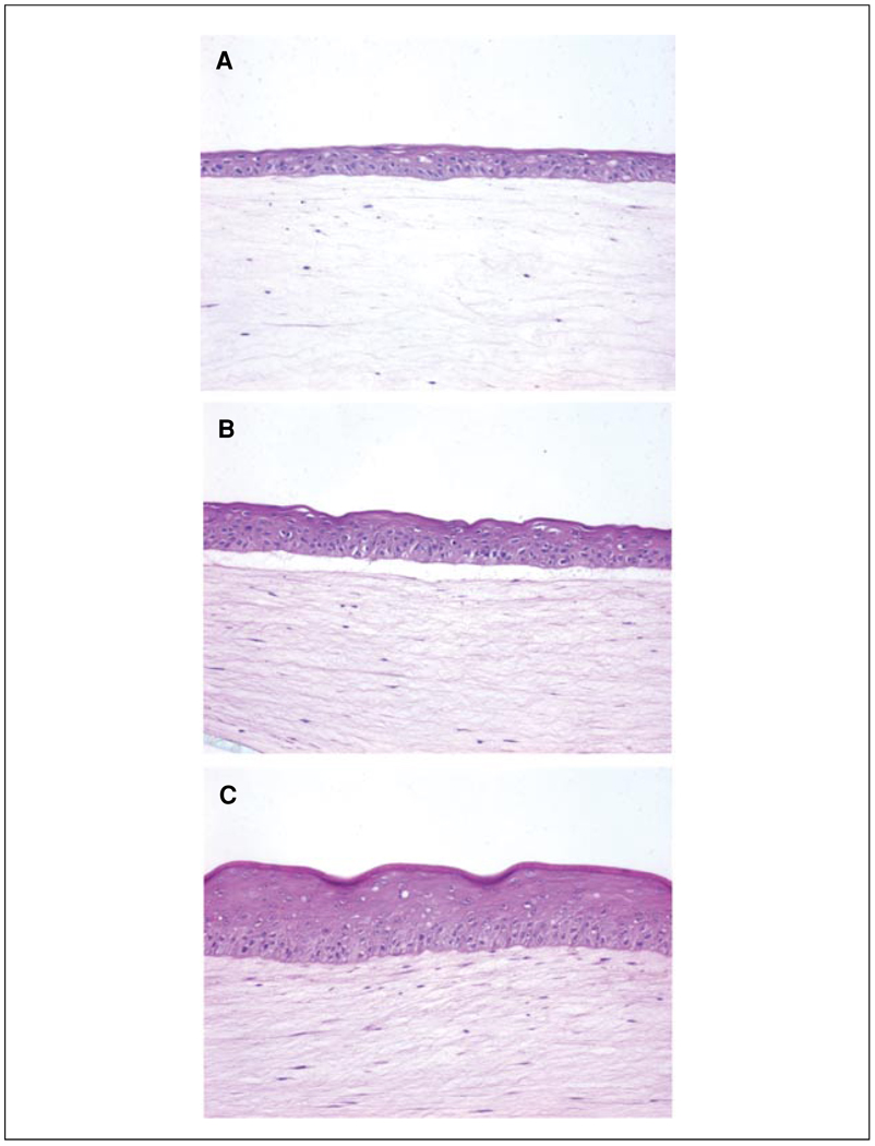
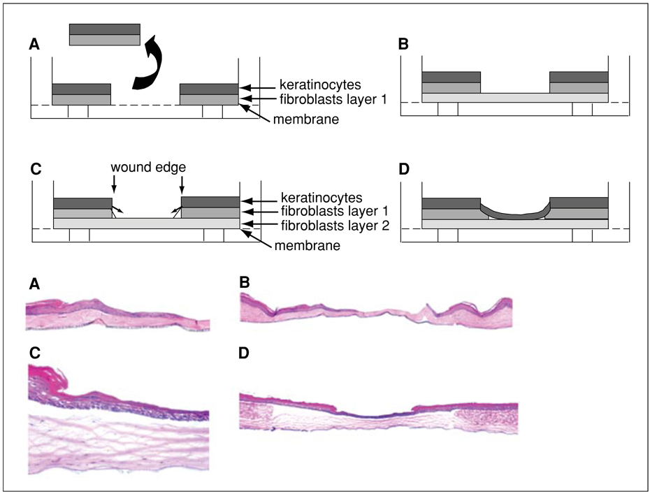
Over the last decade, the development of in vitro, human, three-dimensional (3D) tissue models, known as human skin equivalents (HSEs), has furthered understanding of epidermal cell biology and provided novel experimental systems. Signaling pathways that mediate the linkage between growth and differentiation function optimally when cells are spatially organized to display the architectural features seen in vivo, but are uncoupled and lost in two-dimensional culture systems. HSEs consist of a stratified squamous epithelium grown at an air-liquid interface on a collagen matrix populated with dermal fibroblasts. These 3D tissues demonstrate in vivo-like epithelial differentiation and morphology, and rates of cell division, similar to those found in human skin. This unit describes fabrication of HSEs, allowing the generation of human tissues that mimic the morphology, differentiation, and growth of human skin, as well as disease processes of cancer and wound re-epithelialization, providing powerful new tools for the study of diseases in humans.


Figures

Figure 19.9.1
Figure 19.9.1
Schematic of three-dimensional tissue construction. (A) A thin, acellular layer of collagen is first constructed; it provides an attachment substrate for the cellular collagen. (B) A collagen gel embedded with human dermal fibroblasts is layered onto the acellular layer. (C) While submerged in medium for 7 days, dermal fibroblasts remodel the collagen matrix, causing it to contract away from the walls of the insert. The contracted collagen forms a plateau. (D) Keratinocytes are then added to the center of the plateau of contracted collagen and allowed to attach to the collagen (or intervening substrate such as AlloDerm) to create a monolayer that will form the basal layer of the tissue. (E) Tissues are raised to an air-liquid interface to initiate stratification. Keratinocytes stratify and differentiate and form a suprabasal layer that mimics in vivo skin both morphologically and biochemically. (F) Further exposure to the air-liquid interface and additional feedings with cornification medium results in an increase in the thickness of the spinous and cornified layers of the tissue.
Figure 19.9.2
Figure 19.9.2
Morphologic development of human skin equivalents. Keratinocytes were grown directly on collagen gels for 4 days (A), 6 days (B), and 10 days (C). (A) Early epithelial development, evidenced by a thin epithelium, is seen while tissues are still submerged in epidermalization II medium. (B) The epithelium demonstrates a greater degree of tissue architecture and organization, demonstrated by the presence of cuboidal basal cells, after the tissue is exposed to the air-liquid interface for 2 days. (C) Full morphologic differentiation and stratification are seen after cells are exposed to the air-liquid interface for 6 days. Note that the clear space between the epidermis and dermis in panels A and B likely represents separation along the epithelium-stromal interface due to incomplete organization of the basement membrane at early time points of tissue development. A more mature tissue (C) has a more well developed basement membrane and is more resistant to separation during tissue processing.
Figure 19.9.3
Figure 19.9.3
Three-dimensional tissue model of wound re-epithelialization: schematic (upper panels) and photomicrographs (lower panels; magnification: 5×). (A) A wound is generated through the full thickness of a human skin equivalent (HSE) and the excised tissue is removed. (B) The wounded tissue is placed on a second, contracted collagen gel. (C) Keratinocytes undergo migration to close the wound gap. (D) Keratinocytes have restored epithelial integrity, have closed the wound gap, and undergo stratification.

References

    1. Andriani F, Margulis A, Lin N, Griffey S, Garlick JA. Analysis of microenvironmental factors contributing to basement membrane assembly and normalized epidermal phenotype. J. Invest. Dermatol. 2003;120:923–931. - PubMed
    1. Andriani F, Garfield J, Fusenig NE, Garlick JA. Basement membrane proteins promote progression of intraepithelial neoplasia in 3-dimensional models of human stratified epithelium. Int. J. Cancer. 2004;108:348–357. - PubMed
    1. Bissell MJ, Radisky D. Putting tumours in context. Nat. Rev. Cancer JID. 2001;1011241681:46–54. - PMC - PubMed
    1. Carlson MW, Iyer VR, Marcotte EM. Quantitative gene expression assessment identifies appropriate cell line models for individual cervical cancer pathways. BMC Genomics. 2007;8:117. - PMC - PubMed
    1. Garlick JA, Taichman LB. Effect of TGF-beta 1 on re-epithelialization of human keratinocytes in vitro: An organotypic model. J. Invest. Dermatol. 1994a;103:554–559. - PubMed

LinkOut - more resources