. 2009 Apr;219(1):57-68.
doi: 10.1002/jcp.21646.

TGF-beta promotes cell death and suppresses lactation during the second stage of mammary involution

Affiliations

TGF-beta promotes cell death and suppresses lactation during the second stage of mammary involution

Brian Bierie et al. J Cell Physiol. 2009 Apr.

Abstract

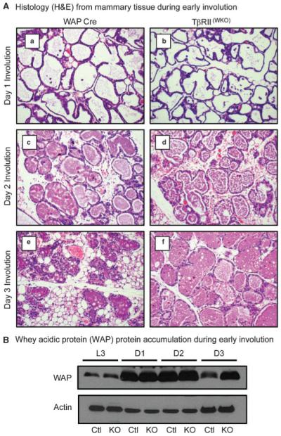
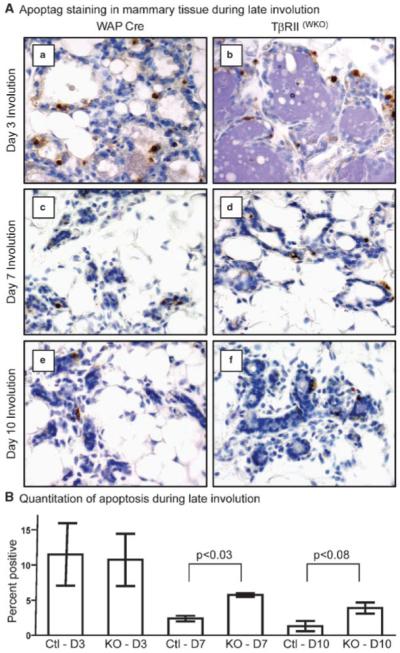
Transforming growth factor beta (TGF-beta) ligands are known to regulate virgin mammary development and contribute to initiation of post-lactation involution. However, the role for TGF-beta during the second phase of mammary involution has not been addressed. Previously, we have used an MMTV-Cre transgene to delete exon 2 from the Tgfbr2 gene in mammary epithelium, however we observed a gradual loss of T beta RII deficient epithelial cells that precluded an accurate study of the role for TGF-beta signaling during involution timepoints. Therefore, in order to determine the role for TGF-beta during the second phase of mammary involution we have now targeted T beta RII ablation within mammary epithelium using the WAP-Cre transgene [T beta RII(WKO)Rosa26R]. Our results demonstrated that TGF-beta regulates commitment to cell death during the second phase of mammary involution. Importantly, at day 3 of mammary involution the Na-Pi type IIb co-transporter (Npt2b), a selective marker for active lactation in luminal lobular alveolar epithelium, was completely silenced in the WAP-Cre control and T beta RII(WKO)Rosa26R tissues. However, by day 7 of involution the T beta RII(WKO)Rosa26R tissues had distended lobular alveoli and regained a robust Npt2b signal that was detected at the apical luminal surface. The Npt2b abundance and localization positively correlated with elevated WAP mRNA expression, suggesting that the distended alveoli were the result of an active lactation program rather than residual milk protein and lipid accumulation. In summary, the results suggest that an epithelial cell response to TGF-beta signaling regulates commitment to cell death and suppression of lactation during the second phase of mammary involution.


Figures

Fig. 1
Fig. 1
Histology and accumulation of whey acidic protein (WAP) during early involution. A: Histology from mammary tissues during involution. a,c,e: Control mammary tissues expressing only the WAP-Cre transgene at days 1 (a), 2 (c), and 3 (e) after forced involution. b,d,f: TβRII(WKO)Rosa26R tissues at days 1 (b), 2 (d), and 3 (f) after forced involution. No differences were noted in histology associated with the first 2 days of involution. However, by the third day of involution control lobular alveolar structures began to collapse while many lobular alveolar structures in TβRII(WKO)Rosa26R tissues at the same time point remained distended. B: Western blot analysis of WAP protein abundance indicated an accumulation in TβRII(WKO)Rosa26R tissues when compared with the control tissues at the same timepoint. Ctl, WAP-Cre; KO, TβRII(WKO)Rosa26R; L3, lactation day 3; D1, involution day 1; D2, involution day 2; D3, involution day 3.
Fig. 2
Fig. 2
Histology during late stages of mammary involution and remodeling. A: Histology of mammary tissue associated with day 7 after forced involution. a,b: Control mammary tissues expressing only the WAP-Cre transgene. c,d: TβRII(WKO)Rosa26R tissues. At low magnification (a,c) the control tissues appeared largely remodeled with minimal evidence of residual terminally differentiated lobular alveolar structures while TβRII(WKO)Rosa26R tissues display an intermediate stage of partial remodeling with many residual expanded lobular alveoli. At higher magnification (b,d), the alveoli in control tissues appear to have returned to a virgin like state while the TβRII(WKO)Rosa26R tissues displayed the presence an eosin stained protein component in addition to abundant lipid droplets within the alveolar lumina. B: Histology of mammary tissue associated with day 10 after forced involution. a,b: Control mammary tissues expressing only the WAP-Cre transgene. c,d: TβRII(WKO)Rosa26R tissues. The control tissue at this timepoint exhibited nearly complete remodeling at low (a) and high magnification (b). TβRII(WKO)Rosa26R tissues at this timepoint remained in an intermediate state of partial involution and remodeling (c). At higher magnification distended alveoli were visible (d), however the expansion was not as prevalent at this timepoint when compared with tissues from day 7 of involution.
Fig. 3
Fig. 3
Apoptosis analysis at days 3, 7, and 10 of involution. A: Apoptag (TUNEL) IHC of mammary tissues during involution. a,c,e: Control mammary tissues expressing only the WAP-Cre transgene at days 3 (a), 7 (c), and 10 (e) after forced involution. b,d,f: TβRII(WKO)Rosa26R tissues at days 3 (b), 7 (d), and 10 (f) after forced involution. B: Quantitation of percent Apoptag positive cells at days 3 (D3), 7 (D7), and 10 (D10) of involution. Three random apoptotic fields per section were counted with three individual mice per genotype at each timepoint. The increased apoptosis detected in the TβRII(WKO)Rosa26R tissues likely reflects the delay of involution whereas this process is nearly complete in the WAP-Cre model by days 7 and 10 of involution.
Fig. 4
Fig. 4
Stat3 and p53 during late stages of mammary involution. A: At day 3 of involution, activated Stat3 (phosphorylation of Tyr-705) was more prevalent in TβRII(WKO)Rosa26R tissues. However, p53 protein abundance was not significantly elevated at this timepoint. Stat3α was more prevalent than Stat3β in both models at this timepoint. β-actin was used as a loading control. Ctl, WAP-Cre; KO, TβRII(WKO)Rosa26R. B: At 7 days of involution, Stat3 activation was comparable between the control and TβRII(WKO)Rosa26R models. Stat3β was observed in both models at this timepoint, however p53 protein abundance was below the threshold of detection by Western blot in both models. β-actin was used as a loading control. C: At day 10 after forced involution, Stat3 activation was significantly reduced in the control and TβRII(WKO)Rosa26R models with only one of the three TβRII(WKO)Rosa26R models exhibiting an elevated level of Stat3 activation at this timepoint. Stat3α and Stat3β levels were comparable in the control and TβRII(WKO)Rosa26R models. p53 protein abundance was below the threshold of detection by Western blot in both models. D: Although p53 protein abundance was below the threshold for Western blot detection, p53 mRNA expression analyses by Northern blot revealed an elevated level of expression in TβRII(WKO)Rosa26R tissues at days 7 and 10 of involution when compared with the WAP-Cre controls. CycA, cyclophillin A was used as a loading control. Ctl, WAP-Cre; KO, TβRII(WKO)Rosa26R.
Fig. 5
Fig. 5
Analysis of Npt2b IF during lactation and early involution. A: IF analysis to determine abundance and localization of Npt2b during lactation. Npt2b was present at the apical surface of lobular alveoli whereas no IF signal was detected in association with ductal epithelium (a, arrow). During lactation, no significant differences were observed when comparing the WAP-Cre (a) and TβRII(WKO)Rosa26R tissues (b). B: IF analysis to determine abundance and localization of Npt2b during days 1 and 2 of involution. No significant difference in Npt2b localization or abundance were observed at either timepoint when comparing WAP-Cre and TβRII(WKO)Rosa26R tissues. In control and TβRII(WKO)Rosa26R tissues no shift in Npt2b IF silencing was observed that would indicate a significant difference in the initiation of the second phase of mammary involution.
Fig. 6
Fig. 6
Presence of a lactogenic phenotype during late stages of mammary involution. A: Npt2b IF detection at day 10 after forced involution. a: WAP-Cre control tissues did not display a Npt2b IF signal, while TβRII(WKO)Rosa26R tissues (b) had a robust Npt2b signal at the apical surface of luminal lobular alveolar epithelial cells. B: Whey acidic protein (WAP) mRNA expression during late involution. WAP expression was completely silenced in WAP-Cre control tissues by day 7 of involution, however it was expressed at a relatively high level in TβRII(WKO)Rosa26R tissues. CypA, cyclophillin A was used as a loading control. C: Localization of Npt2b in virgin TβRII(MKO) and MMTV-DNIIR tissues. Aberrant terminally differentiated lobular alveolar structures obtained from mice during virgin mammary development expressed Npt2b at the luminal apical epithelial surface in both alternate models of mammary epithelial cell specific TGF-β signaling deficiency.

References

    1. Andrechek ER, Hardy WR, Siegel PM, Rudnicki MA, Cardiff RD, Muller WJ. Amplification of the neu/erbB-2 oncogene in a mouse model of mammary tumorigenesis. Proc Natl Acad Sci USA. 2000;97:3444–3449. - PMC - PubMed
    1. Bailey JP, Nieport KM, Herbst MP, Srivastava S, Serra RA, Horseman ND. Prolactin and transforming growth factor-beta signaling exert opposing effects on mammary gland morphogenesis, involution, and the Akt-forkhead pathway. Mol Endocrinol (Baltimore, MD) 2004;18:1171–1184. - PubMed
    1. Bierie B, Stover DG, Abel TW, Chytil A, Gorska AE, Aakre M, Forrester E, Yang L, Wagner KU, Moses HL. Transforming growth factor-beta regulates mammary carcinoma cell survival and interaction with the adjacent microenvironment. Cancer Res. 2008;68:1809–1819. - PubMed
    1. Chapman RS, Lourenco PC, Tonner E, Flint DJ, Selbert S, Takeda K, Akira S, Clarke AR, Watson CJ. Suppression of epithelial apoptosis and delayed mammary gland involution in mice with a conditional knockout of Stat3. Genes Dev. 1999;13:2604–2616. - PMC - PubMed
    1. Chomczynski P, Sacchi N. Single-step method of RNA isolation by acid guanidinium thiocyanate-phenol-chloroform extraction. Anal Biochem. 1987;162:156–159. - PubMed

Publication types

MeSH terms

Substances