. 2009 Mar;63(3):173-80.
doi: 10.1002/syn.20593.

Bursting activation of prefrontal cortex drives sustained up states in nucleus accumbens spiny neurons in vivo

Affiliations

Bursting activation of prefrontal cortex drives sustained up states in nucleus accumbens spiny neurons in vivo

Aaron J Gruber et al. Synapse. 2009 Mar.

Abstract

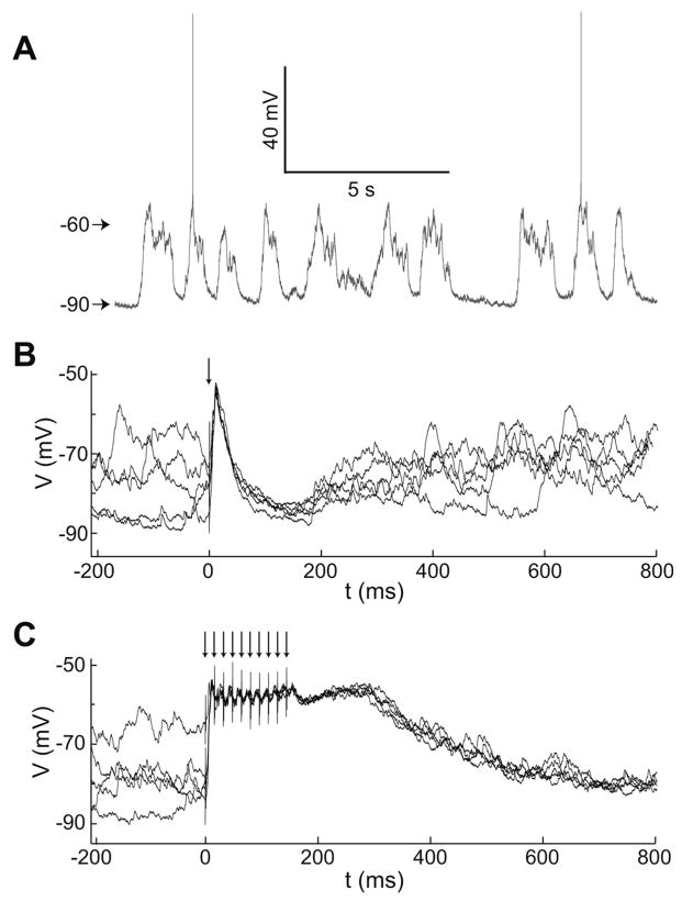
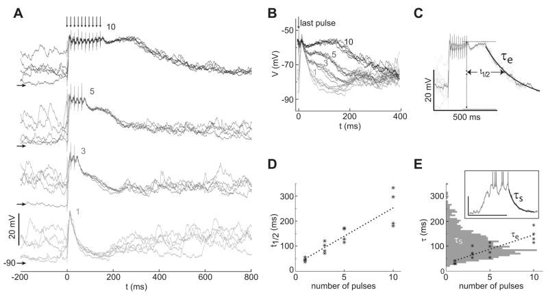
Hippocampal inputs to the nucleus accumbens (NA) have been proposed to implement a gating mechanism by driving NA medium spiny neurons (MSNs) to depolarized up states that facilitate action potential firing in response to brief activation of the prefrontal cortex (PFC). Brief PFC stimulation alone, on the other hand, could not drive NA up states. As these studies were conducted using single-pulse PFC stimulation, it remains possible that PFC activation with naturalistic, bursty patterns can also drive up states in NA MSNs. Here, we assessed NA responses to PFC stimulation with a pattern similar to what is typically observed in awake animals during PFC-relevant behaviors. In vivo intracellular recordings from NA MSNs revealed that brief 20-50 Hz PFC stimulus trains evoked depolarizations that were similar to spontaneous up states in NA MSNs and were sustained beyond stimulus offset. Similar train stimulation of corticoaccumbens afferents in a parasagittal slice preparation evoked large amplitude depolarizations in NA MSNs that were sustained during stimulation but decayed rapidly following stimulation offset, suggesting that activation of cortical afferents can drive MSN depolarizations but other mechanisms may contribute to sustaining up states. These data suggest that NA MSNs integrate temporal features of PFC activation and that the NA gating model can be reformulated to include a PFC-driven gating mechanism during periods of high PFC firing, such as during cognitively demanding tasks. Synapse 63:173-180, 2009. (c) 2008 Wiley-Liss, Inc.


Figures

Fig. 1
Fig. 1
Intracellular recordings of MSNs in vivo. (A) Membrane potential trace from a representative MSN recorded in vivo showing spontaneous fluctuations between hyperpolarized and depolarized potentials. (B) Overlay of traces showing the response of a representative MSN to multiple trials of single-pulse electrical stimulation of PFC. (C) Overlay of traces showing the response of the same MSN as in (B) to multiple trials of PFC stimulation with a 10-pulse train. Arrows indicate times of stimulation pulses in this and in subsequent figures.
Fig. 2
Fig. 2
Dependence of sustained depolarizations on the number of stimulation pulses. (A) Overlay of traces showing the response of a representative MSN to stimulation trains with 1, 3, 5, and 10 pulses (from bottom to top and light to dark traces). Traces are aligned in time to the onset of the first stimulation pulse, and responses to each train length are aligned in the voltage axis (horizontal arrows indicate −90 mV for each response group). (B) The same traces in A aligned to the last stimulation pulse and to a common voltage axis, showing that the trajectory following stimulation offset depends on the number preceding electrical pulses. (C) Illustration of the response features characterized by the time to half decay (t1/2) and decay time constant of the evoked response (τe), superimposed on overlays of the evoked voltage traces showing the response of a MSN to train stimulation. (D) Plot of the time to half decay and the number of pulses for multiple cells revealing a significant linear correlation (n = 4; r2 = 0.81; P < 0.01). (E) Decay time constant (τe) was also correlated with the number of pulses across cells (n = 4; r2 = 0.78; P < 0.01). The graph shows the distribution of decay time constants for the range of pulses tested (asterisks), overlaid with the distribution of decay time constants (horizontal histogram) measured during spontaneous up-down transitions (n = 11). Inset shows an example of τs calculated from an up-down transition.
Fig. 3
Fig. 3
Dependence of sustained depolarizations on stimulation frequency. (A) Overlay of traces from a representative NA neuron showing the subthreshold response to lower frequency stimulation (20 Hz), revealing postsynaptic potentials in response to every pulse in the train. This indicates effective cortico-accumbens transmission throughout the train. (B) Overlay of traces from a representative MSN showing the membrane potential trajectory following stimulation offset for 10-pulse trains at higher frequency (62 Hz, dark traces) and lower frequency (20 Hz, gray trace). (C) Time to half decay is reduced for lower frequency stimulation (n = 4; P < 0.01). (D) Decay time constants are also reduced for lower frequency stimulation (n = 4; P < 0.01).
Fig. 4
Fig. 4
Stimulation of corticostriatal fibers evokes up states in a parasagittal slice preparation. NA spiny neurons were recorded in a whole-cell patch clamp configuration of 0.5–1.0 mm away from the stimulation site along intact corticostriatal fibers. (A) Overlay of voltage responses (top left) to intracellular current pulses (bottom left). Right: I/V plot revealing the characteristic inward rectification in the hyperpolarizing direction. (B) Overlay of average voltage traces recorded in current-clamp mode from a representative NA spiny neuron shows that train stimulation of corticostriatal afferents (arrows) can drive large depolarizations from the hyperpolarized resting down state (dark trace) and that these are greatly attenuated by bath application of the AMPA antagonist CNQX (10 μM; gray trace). (C) Overlay of average voltage traces from a different neuron, showing that depolarization by constant current injection through the recording electrode (upper gray trace) induces a slower repolarization of evoked responses following stimulation offset than responses without such depolarization (lower dark trace). (D) Bar plot showing means and 95% confidence intervals of time to half decay for NA MSN evoked responses in vivo and in the slice preparation with (shaded bars) and without depolarization by current injection through the recording electrode.

References

    1. Bargas J, Howe A, Eberwine J, Surmeier JD. Cellular and molecular characterization of CA2+ currents in acutely isolated. Adult rat neostraial neurons. J Neurosci. 1994;14:6667–6686. - PMC - PubMed
    1. Block AE, Dhanji H, Thompson-Tardif SF, Floresco SB. Thalamic-prefrontal cortical-ventral striatal circuitry mediates dissociable components of strategy set shifting. Cereb Cortex. 2007;17:1625–1636. - PubMed
    1. Bolam JP, Hanley JJ, Booth PA, Bevan MD. Synaptic organization of the basal ganglia. J Anat. 2000;196(Pt 4):527–542. - PMC - PubMed
    1. Brady AM, O’Donnell P. Dopaminergic modulation of prefrontal cortical input to nucleus accumbens neurons in vivo. J Neurosci. 2004;24:1040–1049. - PMC - PubMed
    1. Calabresi P, Pisani A, Mercuri NB, Bernardi G. Long-term potentiation in the striatum is unmasked by removing the voltage-dependent magnesium block of NMDA receptor channels. Eur J Neurosci. 1992;4:929–935. - PubMed

Publication types

LinkOut - more resources